Anna Meyer Children's Hospital, Florence, Italy.
Department of Medical Biotechnology, University of Siena, Siena, Italy.
Pediatr Rheumatol Online J. 2020 Jun 3;18(1):42. doi: 10.1186/s12969-020-00433-w.
Arthritis is often an underestimated extraintestinal manifestation in pediatric inflammatory bowel disease (IBD), including sacroiliitis, whose early signs are well detectable at magnetic resonance imaging (MRI). Magnetic resonance enterography (MRE) is an accurate imaging modality for pediatric IBD assessment. We studied the possibility to detect signs of sacroiliac inflammation in a group of children with IBD who underwent MRE for gastrointestinal disease evaluation.
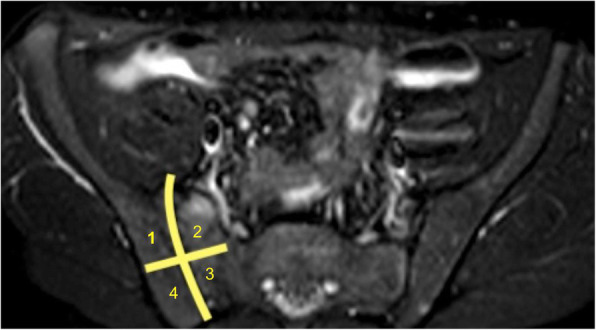
We retrospectively reviewed MRE scans performed in pediatric patients with IBD. We looked for signs of sacroiliitis taking the ASAS (Assessment of SpondyloArthritis international Society) criteria as a model. Presence of bone marrow edema (using T2W sequences with fat suppression), diffusion restriction in Diffusion Weighted Imaging (DWI) or Diffusion Weighted Imaging with Background Suppression (DWIBS), and dynamic contrast enhancement were evaluated. Each SI joint was divided into 4 quadrants: upper iliac, lower iliac, upper sacral, and lower sacral. Two blinded observers with experience in pediatric and skeletal imaging independently evaluated the images. Cases upon which there was a disagreement were evaluated by the two reviewing radiologists and a third radiologist with similar experience together.
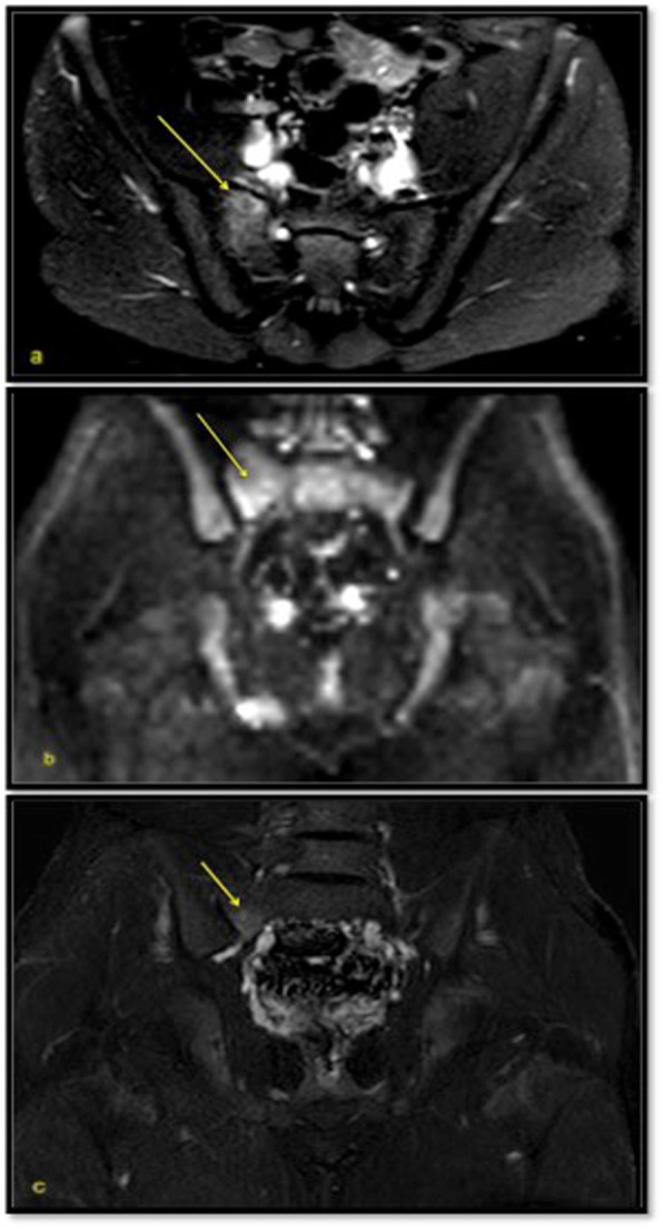
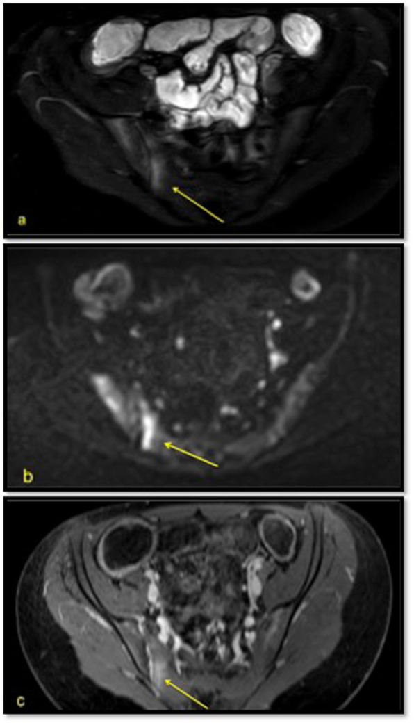
We enrolled 34 patients (24 males and 10 females, with mean age at scanning 14.3 years, median 15.3 years; 2 affected by ulcerative colitis, 32 by Crohn's disease) for a total of 59 examinations performed at the time of their first diagnosis or at symptom exacerbations. No patient complained of musculoskeletal symptoms, neither had pathological findings at articular examination. At the time of MRE 25 patients were under treatment for their IBD. Five patients had radiological signs of SI inflammation at MRE, albeit of mild degree. All patients with SI joint edema also had a restricted diffusion in DWIBS or DWI and almost everyone had contrast media uptake.
Sacroiliitis is one of the extraintestinal manifestation associated with IBD; it is often asymptomatic and clinically underdetected, with an unrelated progression with respect to the underlying IBD. MRE offers the possibility to study SI joints in young patients with IBD who undergo MRE for the investigation of their intestinal condition. Furthermore, we observed that gadolinium enhancement does not improve diagnostic specificity in sacroiliiitis detection.
关节炎是小儿炎症性肠病(IBD)的一种常被低估的肠外表现,包括骶髂关节炎,其早期征象在磁共振成像(MRI)中可很好地检测到。磁共振肠造影术(MRE)是一种用于评估小儿 IBD 的准确成像方式。我们研究了一组接受 MRE 检查以评估胃肠道疾病的 IBD 患儿中检测骶髂关节炎征象的可能性。
我们回顾性分析了接受 MRE 检查的小儿 IBD 患者的 MRE 扫描。我们根据 ASAS(评估脊柱关节炎国际协会)标准作为模型来寻找骶髂关节炎的征象。评估了 T2W 序列(带有脂肪抑制)中的骨髓水肿、弥散受限(在弥散加权成像[DWI]或背景抑制弥散加权成像[DWIBS]中)和动态对比增强。每个骶髂关节分为 4 个象限:髂骨上、髂骨下、骶骨上和骶骨下。两位具有小儿和骨骼成像经验的盲法观察者独立评估图像。存在分歧的病例由两位审查放射科医生和第三位具有类似经验的放射科医生一起评估。
我们共纳入了 34 名患者(24 名男性和 10 名女性,扫描时的平均年龄为 14.3 岁,中位数为 15.3 岁;2 例溃疡性结肠炎,32 例克罗恩病),共进行了 59 次检查,均在首次诊断或症状加重时进行。没有患者出现肌肉骨骼症状,关节检查也无病理发现。在 MRE 时,25 名患者正在接受 IBD 治疗。5 名患者的 MRE 显示骶髂关节炎症的放射学征象,但程度较轻。所有骶髂关节水肿的患者 DWIBS 或 DWI 上均有弥散受限,几乎所有人均有对比剂摄取。
骶髂关节炎是与 IBD 相关的肠外表现之一;它通常无症状,临床漏诊,与潜在的 IBD 无关。MRE 可用于研究因肠道疾病而接受 MRE 检查的年轻 IBD 患者的骶髂关节。此外,我们观察到钆增强并不能提高骶髂关节炎检测的诊断特异性。