• 文献检索
  • 文档翻译
  • 深度研究
  • 学术资讯
  • Suppr Zotero 插件Zotero 插件
  • 邀请有礼
  • 套餐&价格
  • 历史记录
应用&插件
Suppr Zotero 插件Zotero 插件浏览器插件Mac 客户端Windows 客户端微信小程序
定价
高级版会员购买积分包购买API积分包
服务
文献检索文档翻译深度研究API 文档MCP 服务
关于我们
关于 Suppr公司介绍联系我们用户协议隐私条款
关注我们

Suppr 超能文献

核心技术专利:CN118964589B侵权必究
粤ICP备2023148730 号-1Suppr @ 2026

文献检索

告别复杂PubMed语法,用中文像聊天一样搜索,搜遍4000万医学文献。AI智能推荐,让科研检索更轻松。

立即免费搜索

文件翻译

保留排版,准确专业,支持PDF/Word/PPT等文件格式,支持 12+语言互译。

免费翻译文档

深度研究

AI帮你快速写综述,25分钟生成高质量综述,智能提取关键信息,辅助科研写作。

立即免费体验

骶尾部畸胎瘤伴早产:病例报告。

Sacrococcygeal teratoma with preterm delivery: a case report.

机构信息

Gynecology and Obstetrics Department, Corporació Sanitària Parc Taulí, Parc Taulí s/n, 08208, Sabadell, Spain.

出版信息

J Med Case Rep. 2020 Jun 19;14(1):72. doi: 10.1186/s13256-020-02395-9.

DOI:10.1186/s13256-020-02395-9
PMID:32552844
原文链接:https://pmc.ncbi.nlm.nih.gov/articles/PMC7304210/
Abstract

BACKGROUND

Sacrococcygeal teratoma is one of the most frequently prenatally diagnosed neoplasias. Obstetric ultrasound has a role in the diagnosis and management of these tumors during pregnancy. In this report, we describe a multidisciplinary approach in a case of a patient with sacrococcygeal teratomas and preterm delivery, as well as postnatal outcomes.

CASE PRESENTATION

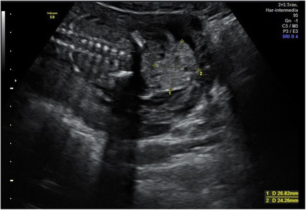
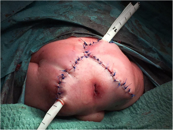
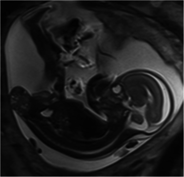
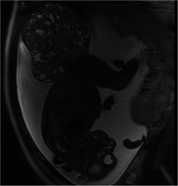
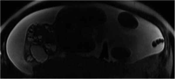
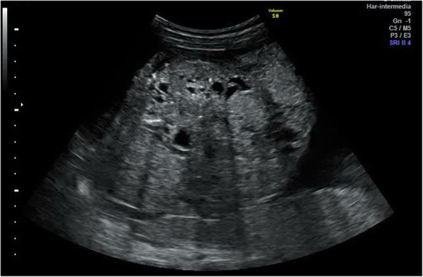
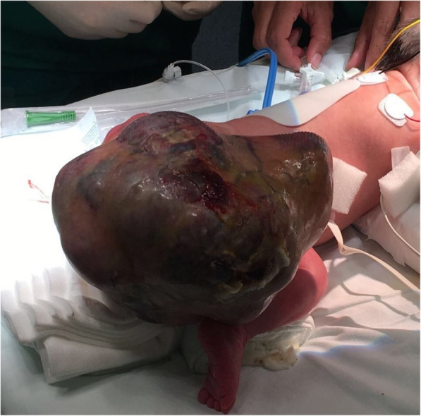
A 26-year-old Caucasian woman at 20.3 weeks of gestation with a normal gestational course and no relevant medical or surgical history was referred to our institution with a sacrococcygeal mass diagnosis. Magnetic resonance imaging confirmed the diagnosis of sacrococcygeal teratoma type I according to the Altman classification. Follow-up with ultrasound showed an increase in the size of the mass up to 190 × 150 mm, high Doppler flow, and severe polyhydramnios. At 35.1 weeks of gestation, the patient had premature rupture of membranes, and an emergency cesarean section was performed due to recurrent late decelerations detected by fetal heart rate monitoring. Afterward, surgery was performed successfully at 36 hours of life. Posterior controls revealed normal and healthy child growth.

CONCLUSIONS

This case report demonstrates the importance of a multidisciplinary approach to offer the best neonatal outcomes by performing early surgery, as well as the need for follow-up by ultrasound in order to minimize complications by assessing mass growth, Doppler flow, and amniotic fluid.

摘要

背景

骶尾部畸胎瘤是最常被产前诊断出的肿瘤之一。产科超声在这些肿瘤的诊断和孕期管理中具有重要作用。本报告描述了一例骶尾部畸胎瘤合并早产患者的多学科治疗方法,以及产后结局。

病例介绍

一名 26 岁的白人女性,妊娠 20.3 周,正常妊娠,无相关内科或外科病史,因骶尾部肿块就诊于我院。磁共振成像(MRI)根据 Altman 分类证实了骶尾部畸胎瘤 I 型的诊断。超声随访显示肿块大小增加至 190×150mm,多普勒血流高,羊水过多。妊娠 35.1 周时,患者胎膜早破,因胎儿心率监测发现晚期减速反复发作,行急诊剖宫产术。随后,患儿出生后 36 小时成功进行了手术。后期随访显示患儿生长正常,健康。

结论

本病例报告强调了多学科治疗的重要性,通过早期手术可以获得最佳的新生儿结局,同时需要通过超声进行随访,以通过评估肿块生长、多普勒血流和羊水来最小化并发症。

https://cdn.ncbi.nlm.nih.gov/pmc/blobs/474f/7304210/873fb6106b36/13256_2020_2395_Fig7_HTML.jpg
https://cdn.ncbi.nlm.nih.gov/pmc/blobs/474f/7304210/fc0fecc2dd7c/13256_2020_2395_Fig1_HTML.jpg
https://cdn.ncbi.nlm.nih.gov/pmc/blobs/474f/7304210/8501f9a0920a/13256_2020_2395_Fig2_HTML.jpg
https://cdn.ncbi.nlm.nih.gov/pmc/blobs/474f/7304210/3414630f8187/13256_2020_2395_Fig3_HTML.jpg
https://cdn.ncbi.nlm.nih.gov/pmc/blobs/474f/7304210/4e1b57731809/13256_2020_2395_Fig4_HTML.jpg
https://cdn.ncbi.nlm.nih.gov/pmc/blobs/474f/7304210/efc6dd6d49a3/13256_2020_2395_Fig5_HTML.jpg
https://cdn.ncbi.nlm.nih.gov/pmc/blobs/474f/7304210/69af562ef299/13256_2020_2395_Fig6_HTML.jpg
https://cdn.ncbi.nlm.nih.gov/pmc/blobs/474f/7304210/873fb6106b36/13256_2020_2395_Fig7_HTML.jpg

相似文献

1
Sacrococcygeal teratoma with preterm delivery: a case report.骶尾部畸胎瘤伴早产:病例报告。
J Med Case Rep. 2020 Jun 19;14(1):72. doi: 10.1186/s13256-020-02395-9.
2
Sacrococcygeal teratoma in one twin: a case report and literature review.骶尾部畸胎瘤一胎内同时发生:病例报告及文献复习。
BMC Pregnancy Childbirth. 2020 Dec 2;20(1):751. doi: 10.1186/s12884-020-03454-1.
3
[Prenatal diagnosis and prognostic factors analysis of fetal sacrococcygeal teratoma].[胎儿骶尾部畸胎瘤的产前诊断及预后因素分析]
Zhonghua Fu Chan Ke Za Zhi. 2022 Jun 25;57(6):413-418. doi: 10.3760/cma.j.cn112141-20220115-00025.
4
Prenatal diagnosis of type I sacrococcygeal teratoma and its management.I型骶尾部畸胎瘤的产前诊断及其处理
Clin Exp Obstet Gynecol. 2008;35(2):153-5.
5
Prenatal identification of a completely cystic internal sacrococcygeal teratoma (type IV).完全囊性骶尾部畸胎瘤(IV型)的产前诊断
Ultrasound Obstet Gynecol. 1997 Jun;9(6):425-8. doi: 10.1046/j.1469-0705.1997.09060425.x.
6
Rapid growth of a fetal sacrococcygeal teratoma in an HIV-infected woman: a case report.一名感染艾滋病毒女性胎儿骶尾部畸胎瘤的快速生长:病例报告
J Reprod Med. 2006 May;51(5):431-4.
7
Sacrococcygeal teratoma: prenatal assessment, fetal intervention, and outcome.骶尾部畸胎瘤:产前评估、胎儿干预及结局
J Pediatr Surg. 2004 Mar;39(3):430-8; discussion 430-8. doi: 10.1016/j.jpedsurg.2003.11.005.
8
Experience with 8 cases of prenatally diagnosed sacrococcygeal teratomas.8例产前诊断的骶尾部畸胎瘤的经验。
Fetal Ther. 1987;2(2):88-94. doi: 10.1159/000263289.
9
Sacrococcygeal teratoma with hydrops fetalis and bilateral hydronephrosis.骶尾部畸胎瘤合并胎儿水肿及双侧肾积水。
J Perinat Med. 2000;28(5):414-8. doi: 10.1515/JPM.2000.054.
10
Three-dimensional power Doppler sonography in the diagnosis of a cystic sacrococcygeal teratoma mimicking a meningomyelocele: A case report.三维能量多普勒超声在诊断酷似脊髓脊膜膨出的骶尾部囊性畸胎瘤中的应用:一例报告
J Clin Ultrasound. 2009 Sep;37(7):410-3. doi: 10.1002/jcu.20592.

引用本文的文献

1
Perinatal Outcomes of Intrauterine Interventions for Fetal Sacrococcygeal Teratoma Based on Different Surgical Techniques-A Systematic Review.基于不同手术技术的胎儿骶尾部畸胎瘤宫内干预围产期结局——一项系统评价
J Clin Med. 2024 Apr 30;13(9):2649. doi: 10.3390/jcm13092649.
2
Transient Polyhydramnios during Pregnancy Complicated with Gestational Diabetes Mellitus: Case Report and Systematic Review.妊娠合并妊娠期糖尿病时的短暂性羊水过多:病例报告及系统评价
Diagnostics (Basel). 2022 May 28;12(6):1340. doi: 10.3390/diagnostics12061340.

本文引用的文献

1
Diagnosis of sacrococcygeal teratoma using two and three-dimensional ultrasonography: two cases reported and a literature review.二维和三维超声检查在骶尾部畸胎瘤诊断中的应用:两例报告并文献复习
Med Ultrason. 2014 Sep;16(3):274-7. doi: 10.11152/mu.2013.2066.163.mg1gi2.
2
Early delivery as an alternative management strategy for selected high-risk fetal sacrococcygeal teratomas.对于选择的高危胎儿骶尾部畸胎瘤,早期分娩是一种替代管理策略。
J Pediatr Surg. 2011 Jul;46(7):1325-32. doi: 10.1016/j.jpedsurg.2010.10.020.
3
Sacrococcygeal teratoma over two decades: birth prevalence, prenatal diagnosis and clinical outcomes.
二十多年来的骶尾部畸胎瘤:出生患病率、产前诊断及临床结局
Prenat Diagn. 2008 Nov;28(11):1048-51. doi: 10.1002/pd.2122.
4
Management and outcome in prenatally diagnosed sacrococcygeal teratomas.产前诊断的骶尾部畸胎瘤的管理与预后
Pediatr Int. 2008 Aug;50(4):576-80. doi: 10.1111/j.1442-200X.2008.02703.x.
5
[Neonatal neoplasms: a single-centre experience].[新生儿肿瘤:单中心经验]
An Pediatr (Barc). 2007 Jul;67(1):85-6. doi: 10.1157/13108090.
6
Terminal myelocystocele and sacrococcygeal teratoma: a comparison of fetal ultrasound presentation and perinatal risk.终末脊髓脊膜膨出与骶尾部畸胎瘤:胎儿超声表现及围产期风险比较
AJNR Am J Neuroradiol. 2007 Jun-Jul;28(6):1058-60. doi: 10.3174/ajnr.A0502.
7
Prenatally diagnosed sacrococcygeal teratoma: a prognostic classification.产前诊断的骶尾部畸胎瘤:一种预后分类。
J Pediatr Surg. 2006 Sep;41(9):1517-21. doi: 10.1016/j.jpedsurg.2006.05.009.
8
Sacrococcygeal teratoma--a 25-year experience in a UK regional center.骶尾部畸胎瘤——英国某地区中心25年的经验
J Pediatr Surg. 2006 Sep;41(9):1513-6. doi: 10.1016/j.jpedsurg.2006.05.019.
9
Perinatal (fetal and neonatal) germ cell tumors.围产期(胎儿期和新生儿期)生殖细胞肿瘤。
J Pediatr Surg. 2004 Jul;39(7):1003-13. doi: 10.1016/j.jpedsurg.2004.03.045.
10
Sacrococcygeal teratoma in the perinatal period.围产期骶尾部畸胎瘤。
Postgrad Med J. 2000 Dec;76(902):754-9. doi: 10.1136/pmj.76.902.754.