• 文献检索
  • 文档翻译
  • 深度研究
  • 学术资讯
  • Suppr Zotero 插件Zotero 插件
  • 邀请有礼
  • 套餐&价格
  • 历史记录
应用&插件
Suppr Zotero 插件Zotero 插件浏览器插件Mac 客户端Windows 客户端微信小程序
定价
高级版会员购买积分包购买API积分包
服务
文献检索文档翻译深度研究API 文档MCP 服务
关于我们
关于 Suppr公司介绍联系我们用户协议隐私条款
关注我们

Suppr 超能文献

核心技术专利:CN118964589B侵权必究
粤ICP备2023148730 号-1Suppr @ 2026

文献检索

告别复杂PubMed语法,用中文像聊天一样搜索,搜遍4000万医学文献。AI智能推荐,让科研检索更轻松。

立即免费搜索

文件翻译

保留排版,准确专业,支持PDF/Word/PPT等文件格式,支持 12+语言互译。

免费翻译文档

深度研究

AI帮你快速写综述,25分钟生成高质量综述,智能提取关键信息,辅助科研写作。

立即免费体验

MLKL 在质膜上的转运和聚集控制着细胞发生坏死性凋亡的动力学和阈值。

MLKL trafficking and accumulation at the plasma membrane control the kinetics and threshold for necroptosis.

机构信息

The Walter and Eliza Hall Institute of Medical Research, Parkville, VIC, Australia.

Department of Medical Biology, The University of Melbourne, Parkville, VIC, Australia.

出版信息

Nat Commun. 2020 Jun 19;11(1):3151. doi: 10.1038/s41467-020-16887-1.

DOI:10.1038/s41467-020-16887-1
PMID:32561730
原文链接:https://pmc.ncbi.nlm.nih.gov/articles/PMC7305196/
Abstract

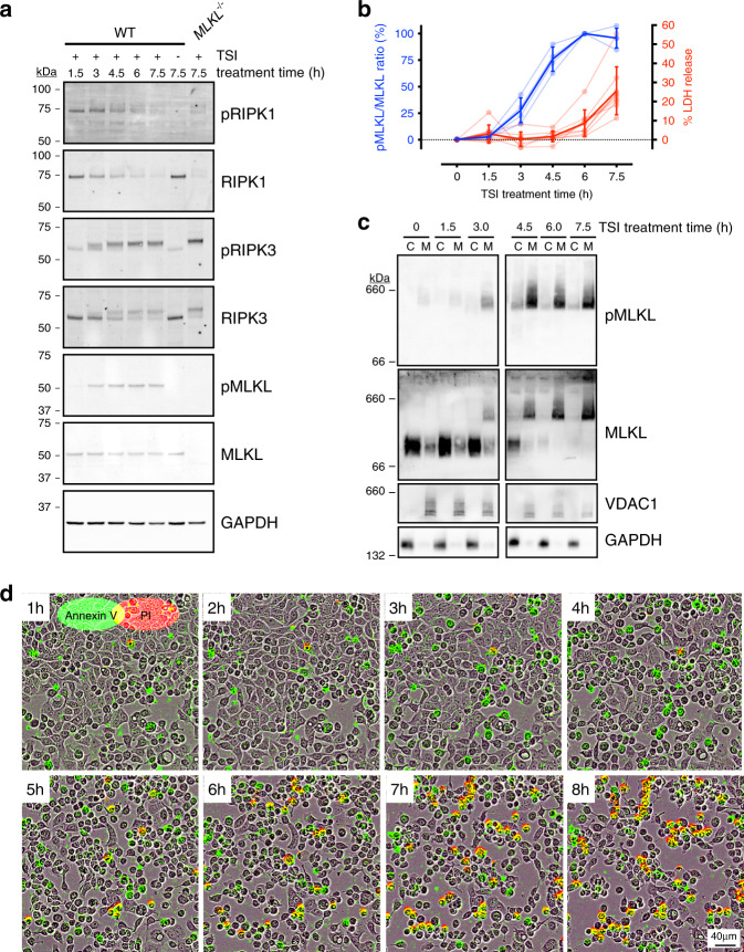
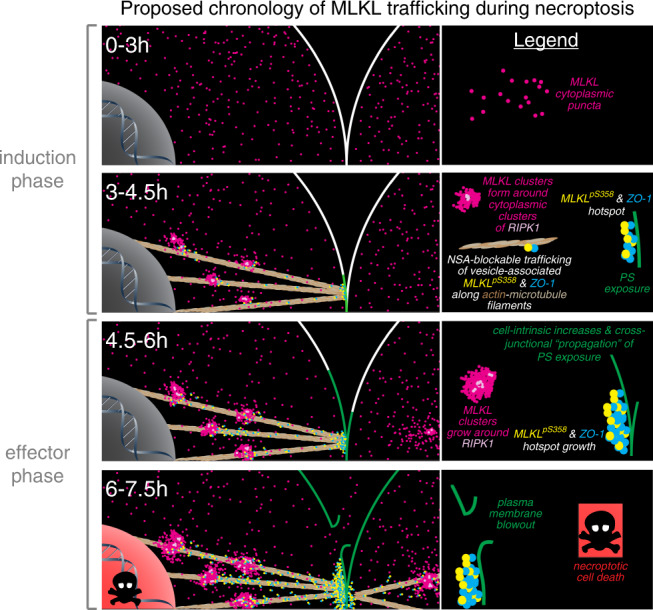
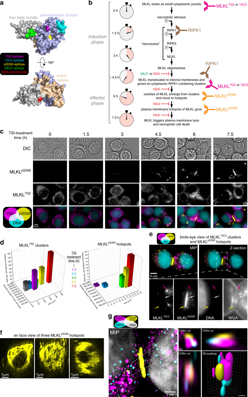
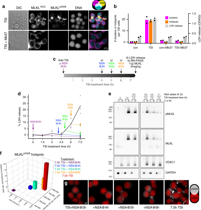
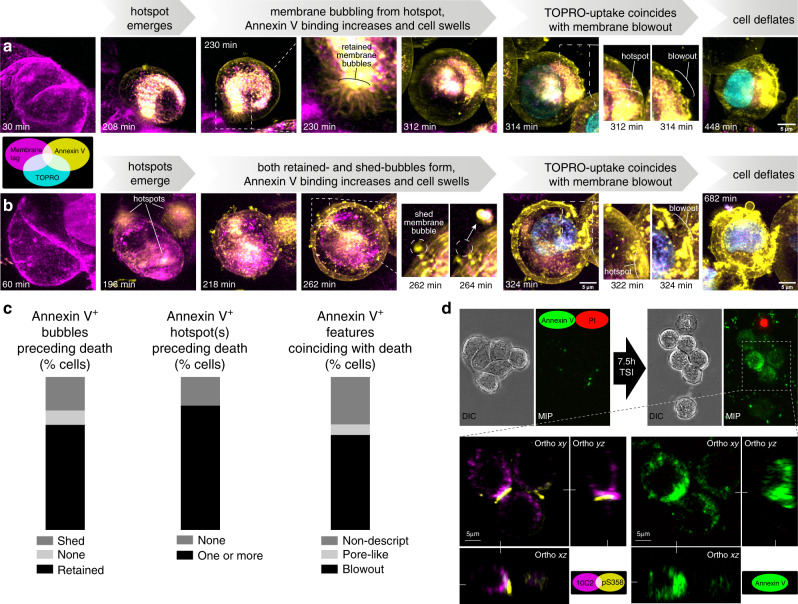
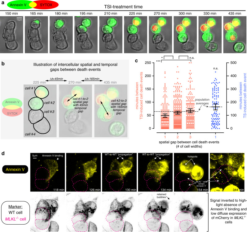
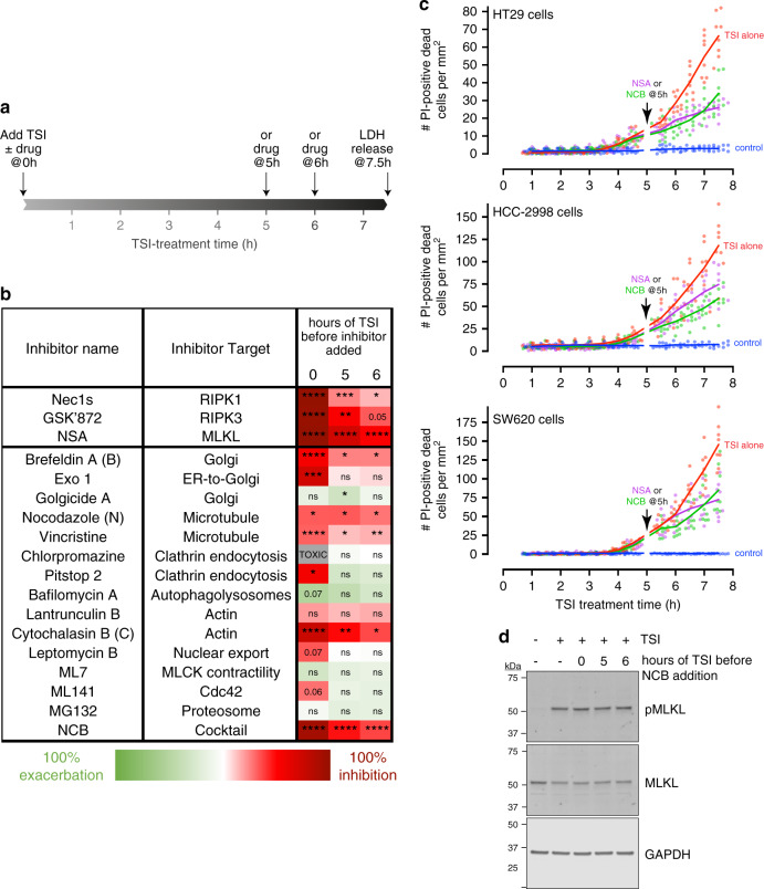
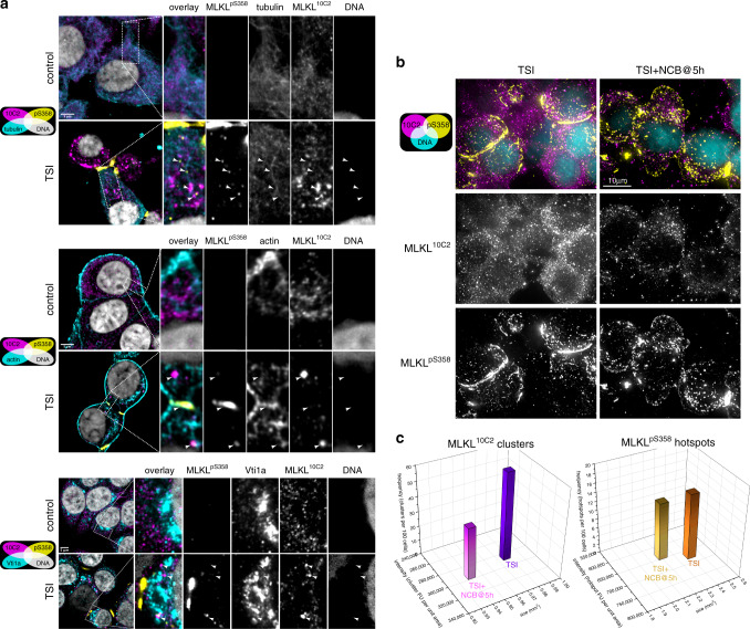
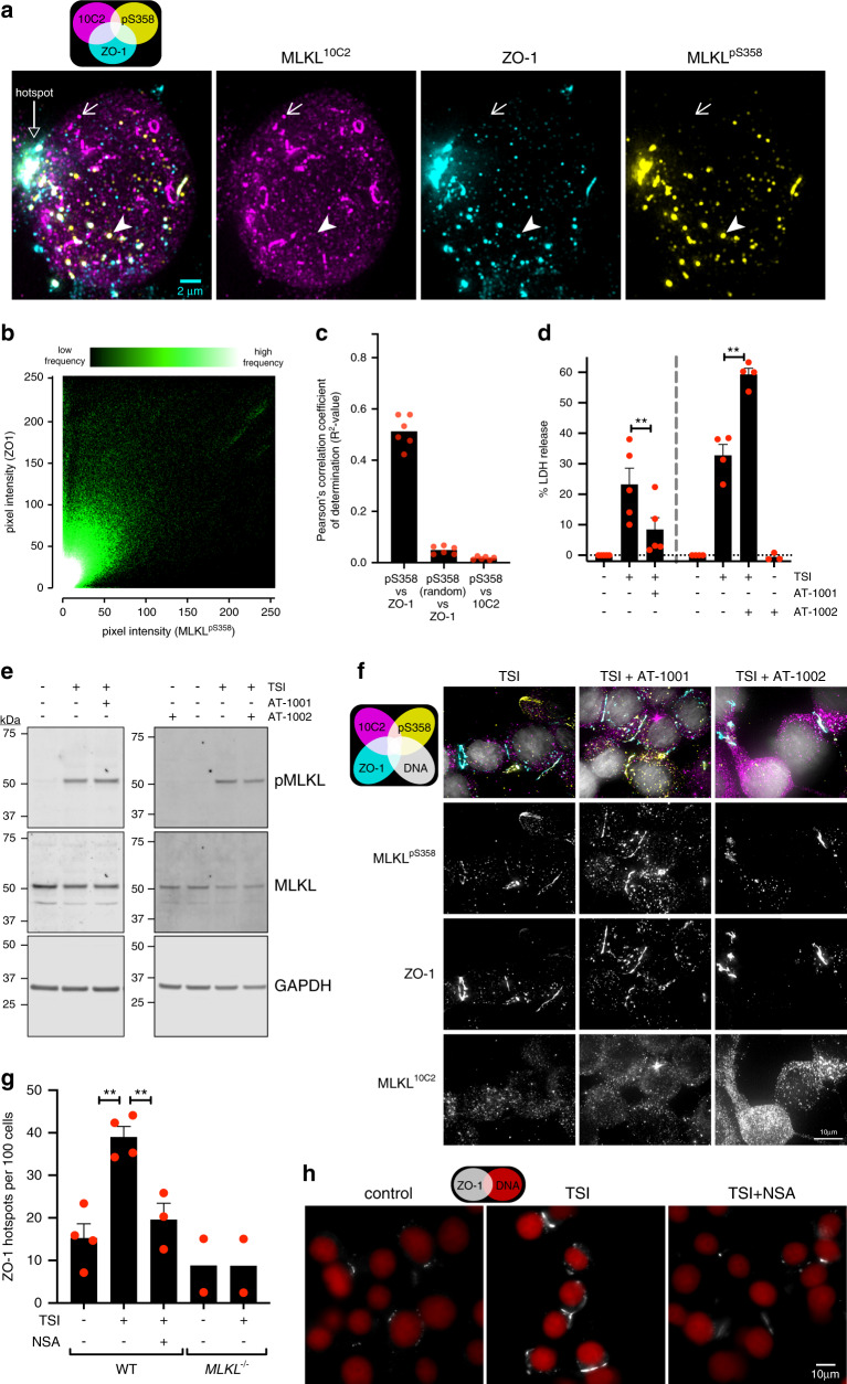
Mixed lineage kinase domain-like (MLKL) is the terminal protein in the pro-inflammatory necroptotic cell death program. RIPK3-mediated phosphorylation is thought to initiate MLKL oligomerization, membrane translocation and membrane disruption, although the precise choreography of events is incompletely understood. Here, we use single-cell imaging approaches to map the chronology of endogenous human MLKL activation during necroptosis. During the effector phase of necroptosis, we observe that phosphorylated MLKL assembles into higher order species on presumed cytoplasmic necrosomes. Subsequently, MLKL co-traffics with tight junction proteins to the cell periphery via Golgi-microtubule-actin-dependent mechanisms. MLKL and tight junction proteins then steadily co-accumulate at the plasma membrane as heterogeneous micron-sized hotspots. Our studies identify MLKL trafficking and plasma membrane accumulation as crucial necroptosis checkpoints. Furthermore, the accumulation of phosphorylated MLKL at intercellular junctions accelerates necroptosis between neighbouring cells, which may be relevant to inflammatory bowel disease and other necroptosis-mediated enteropathies.

摘要

混合谱系激酶结构域样(MLKL)是促炎坏死性细胞死亡程序中的末端蛋白。RIPK3 介导的磷酸化被认为是 MLKL 寡聚化、膜易位和膜破坏的起始事件,尽管事件的确切顺序尚不完全清楚。在这里,我们使用单细胞成像方法来绘制内源性人 MLKL 在坏死性细胞死亡过程中的激活顺序。在坏死性细胞死亡的效应阶段,我们观察到磷酸化的 MLKL 在假定的细胞质坏死小体上组装成更高阶的物种。随后,MLKL 通过高尔基体-微管-肌动蛋白依赖的机制与紧密连接蛋白一起运输到细胞外周。MLKL 和紧密连接蛋白随后作为异质的微米大小热点稳定地在质膜上积累。我们的研究确定了 MLKL 的运输和质膜积累是坏死性细胞死亡的关键检查点。此外,磷酸化 MLKL 在细胞间连接点的积累加速了相邻细胞之间的坏死性细胞死亡,这可能与炎症性肠病和其他坏死性细胞死亡介导的肠病有关。

https://cdn.ncbi.nlm.nih.gov/pmc/blobs/71d8/7305196/e05604eaffc1/41467_2020_16887_Fig9_HTML.jpg
https://cdn.ncbi.nlm.nih.gov/pmc/blobs/71d8/7305196/6dcf27ec1075/41467_2020_16887_Fig1_HTML.jpg
https://cdn.ncbi.nlm.nih.gov/pmc/blobs/71d8/7305196/2a79a54beabb/41467_2020_16887_Fig2_HTML.jpg
https://cdn.ncbi.nlm.nih.gov/pmc/blobs/71d8/7305196/cd7a5a95a07f/41467_2020_16887_Fig3_HTML.jpg
https://cdn.ncbi.nlm.nih.gov/pmc/blobs/71d8/7305196/a99520440d0d/41467_2020_16887_Fig4_HTML.jpg
https://cdn.ncbi.nlm.nih.gov/pmc/blobs/71d8/7305196/7be2a3f75d24/41467_2020_16887_Fig5_HTML.jpg
https://cdn.ncbi.nlm.nih.gov/pmc/blobs/71d8/7305196/3f9290ba3914/41467_2020_16887_Fig6_HTML.jpg
https://cdn.ncbi.nlm.nih.gov/pmc/blobs/71d8/7305196/fbd6df2d3b70/41467_2020_16887_Fig7_HTML.jpg
https://cdn.ncbi.nlm.nih.gov/pmc/blobs/71d8/7305196/8f2259c2304b/41467_2020_16887_Fig8_HTML.jpg
https://cdn.ncbi.nlm.nih.gov/pmc/blobs/71d8/7305196/e05604eaffc1/41467_2020_16887_Fig9_HTML.jpg

相似文献

1
MLKL trafficking and accumulation at the plasma membrane control the kinetics and threshold for necroptosis.MLKL 在质膜上的转运和聚集控制着细胞发生坏死性凋亡的动力学和阈值。
Nat Commun. 2020 Jun 19;11(1):3151. doi: 10.1038/s41467-020-16887-1.
2
A toolbox for imaging RIPK1, RIPK3, and MLKL in mouse and human cells.用于在小鼠和人细胞中成像 RIPK1、RIPK3 和 MLKL 的工具包。
Cell Death Differ. 2021 Jul;28(7):2126-2144. doi: 10.1038/s41418-021-00742-x. Epub 2021 Feb 15.
3
The NS1 Protein of Influenza A Virus Participates in Necroptosis by Interacting with MLKL and Increasing Its Oligomerization and Membrane Translocation.甲型流感病毒 NS1 蛋白通过与 MLKL 相互作用并增加其寡聚化和膜转位来参与坏死性凋亡。
J Virol. 2019 Jan 4;93(2). doi: 10.1128/JVI.01835-18. Print 2019 Jan 15.
4
Identification of MLKL membrane translocation as a checkpoint in necroptotic cell death using Monobodies.利用单域抗体鉴定 MLKL 膜易位作为坏死性细胞死亡的检查点。
Proc Natl Acad Sci U S A. 2020 Apr 14;117(15):8468-8475. doi: 10.1073/pnas.1919960117. Epub 2020 Mar 31.
5
Conformational interconversion of MLKL and disengagement from RIPK3 precede cell death by necroptosis.MLKL 构象转换并与 RIPK3 脱离后,细胞发生坏死性细胞死亡。
Nat Commun. 2021 Apr 13;12(1):2211. doi: 10.1038/s41467-021-22400-z.
6
Translocation of mixed lineage kinase domain-like protein to plasma membrane leads to necrotic cell death.混合谱系激酶结构域样蛋白易位至质膜导致坏死性细胞死亡。
Cell Res. 2014 Jan;24(1):105-21. doi: 10.1038/cr.2013.171. Epub 2013 Dec 24.
7
Characterization of RIPK3-mediated phosphorylation of the activation loop of MLKL during necroptosis.坏死性凋亡过程中RIPK3介导的MLKL激活环磷酸化的特征分析。
Cell Death Differ. 2016 Jan;23(1):76-88. doi: 10.1038/cdd.2015.70. Epub 2015 May 29.
8
LUBAC-mediated M1 Ub regulates necroptosis by segregating the cellular distribution of active MLKL.LUBAC 介导的 M1 Ub 通过分隔活性 MLKL 的细胞分布来调节细胞坏死。
Cell Death Dis. 2024 Jan 20;15(1):77. doi: 10.1038/s41419-024-06447-6.
9
Reconstitution of Human Necrosome Interactions in .在. 中重建人坏死小体相互作用。
Biomolecules. 2021 Jan 25;11(2):153. doi: 10.3390/biom11020153.
10
Diverse sequence determinants control human and mouse receptor interacting protein 3 (RIP3) and mixed lineage kinase domain-like (MLKL) interaction in necroptotic signaling.多种序列决定因素控制人类和小鼠受体相互作用蛋白 3(RIP3)和混合谱系激酶结构域样(MLKL)在坏死性信号转导中的相互作用。
J Biol Chem. 2013 Jun 7;288(23):16247-16261. doi: 10.1074/jbc.M112.435545. Epub 2013 Apr 23.

引用本文的文献

1
Regulated cell death in fungi from a comparative immunology perspective.从比较免疫学角度看真菌中的程序性细胞死亡
Cell Death Differ. 2025 Sep 3. doi: 10.1038/s41418-025-01570-z.
2
Dual role mechanisms of regulated cell death in apical periodontitis: from pathogenic destruction to therapeutic potential.根尖周炎中程序性细胞死亡的双重作用机制:从致病破坏到治疗潜力
Cell Death Discov. 2025 Aug 15;11(1):386. doi: 10.1038/s41420-025-02686-4.
3
Mechanistic Insights into Autophagy-Dependent Cell Death (ADCD): A Novel Avenue for Cancer Therapy.

本文引用的文献

1
Identification of MLKL membrane translocation as a checkpoint in necroptotic cell death using Monobodies.利用单域抗体鉴定 MLKL 膜易位作为坏死性细胞死亡的检查点。
Proc Natl Acad Sci U S A. 2020 Apr 14;117(15):8468-8475. doi: 10.1073/pnas.1919960117. Epub 2020 Mar 31.
2
Necroptosis-blocking compound NBC1 targets heat shock protein 70 to inhibit MLKL polymerization and necroptosis.坏死性凋亡阻断化合物 NBC1 靶向热休克蛋白 70 以抑制 MLKL 聚合和坏死性凋亡。
Proc Natl Acad Sci U S A. 2020 Mar 24;117(12):6521-6530. doi: 10.1073/pnas.1916503117. Epub 2020 Mar 10.
3
Knocking 'em Dead: Pore-Forming Proteins in Immune Defense.
自噬依赖性细胞死亡(ADCD)的机制性见解:癌症治疗的新途径。
Cells. 2025 Jul 13;14(14):1072. doi: 10.3390/cells14141072.
4
Disrupting membranes, controlling cell fate: the role of pore-forming proteins in cell death and therapy.破坏细胞膜,控制细胞命运:成孔蛋白在细胞死亡和治疗中的作用
Apoptosis. 2025 Jul 21. doi: 10.1007/s10495-025-02133-w.
5
Pathogenesis and treatment strategies of sepsis-induced myocardial injury: modern and traditional medical perspectives.脓毒症诱导的心肌损伤的发病机制及治疗策略:现代医学与传统医学视角
Int J Biol Sci. 2025 May 15;21(8):3478-3504. doi: 10.7150/ijbs.111288. eCollection 2025.
6
MLKL‒OPTN axis regulates herpesvirus-induced neurological sequelae.混合谱系激酶结构域样蛋白(MLKL)‒视神经萎缩蛋白(OPTN)轴调控疱疹病毒诱导的神经后遗症。
Clin Transl Med. 2025 Jun;15(6):e70353. doi: 10.1002/ctm2.70353.
7
CO confers neuroprotection via activating the PERK-calcineurin pathway and inhibiting necroptosis.一氧化碳通过激活PERK-钙调神经磷酸酶途径和抑制坏死性凋亡发挥神经保护作用。
Cell Death Discov. 2025 May 27;11(1):254. doi: 10.1038/s41420-025-02530-9.
8
A viral necrosome mediates direct RIPK3 activation to promote inflammatory necroptosis.病毒坏死小体介导RIPK3直接激活以促进炎性坏死性凋亡。
Proc Natl Acad Sci U S A. 2025 Jun 3;122(22):e2420245122. doi: 10.1073/pnas.2420245122. Epub 2025 May 27.
9
Mesenchymal stem cells protect the integrity of the alveolar epithelial barrier through extracellular vesicles by inhibiting MAPK-mediated necroptosis.间充质干细胞通过细胞外囊泡抑制丝裂原活化蛋白激酶(MAPK)介导的坏死性凋亡,从而保护肺泡上皮屏障的完整性。
Stem Cell Res Ther. 2025 May 19;16(1):250. doi: 10.1186/s13287-025-04388-1.
10
Epigenetic Inactivation of RIPK3-Dependent Necroptosis Augments Cisplatin Chemoresistance in Human Osteosarcoma.RIPK3 依赖性坏死性凋亡的表观遗传失活增强了人骨肉瘤对顺铂的化疗耐药性。
Int J Mol Sci. 2025 Apr 18;26(8):3863. doi: 10.3390/ijms26083863.
《一击必杀:免疫防御中的成孔蛋白》
Annu Rev Immunol. 2020 Apr 26;38:455-485. doi: 10.1146/annurev-immunol-111319-023800. Epub 2020 Jan 31.
4
Mutations that prevent caspase cleavage of RIPK1 cause autoinflammatory disease.阻止半胱天冬酶切割 RIPK1 的突变会导致自身炎症性疾病。
Nature. 2020 Jan;577(7788):103-108. doi: 10.1038/s41586-019-1828-5. Epub 2019 Dec 11.
5
A dominant autoinflammatory disease caused by non-cleavable variants of RIPK1.一种由 RIPK1 的不可切割变异体引起的显性自身炎症性疾病。
Nature. 2020 Jan;577(7788):109-114. doi: 10.1038/s41586-019-1830-y. Epub 2019 Dec 11.
6
Viral MLKL Homologs Subvert Necroptotic Cell Death by Sequestering Cellular RIPK3.病毒 MLKL 同源物通过隔离细胞 RIPK3 来颠覆细胞程序性坏死。
Cell Rep. 2019 Sep 24;28(13):3309-3319.e5. doi: 10.1016/j.celrep.2019.08.055.
7
K63-linked ubiquitination regulates RIPK1 kinase activity to prevent cell death during embryogenesis and inflammation.K63 连接的泛素化调节 RIPK1 激酶活性,以防止胚胎发生和炎症期间的细胞死亡。
Nat Commun. 2019 Sep 13;10(1):4157. doi: 10.1038/s41467-019-12033-8.
8
Cleavage of RIPK1 by caspase-8 is crucial for limiting apoptosis and necroptosis.Caspase-8 介导的 RIPK1 切割对于限制细胞凋亡和坏死性凋亡至关重要。
Nature. 2019 Oct;574(7778):428-431. doi: 10.1038/s41586-019-1548-x. Epub 2019 Sep 11.
9
Pannexin-1 limits the production of proinflammatory cytokines during necroptosis.Pannexin-1 在细胞坏死性凋亡过程中限制促炎细胞因子的产生。
EMBO Rep. 2019 Oct 4;20(10):e47840. doi: 10.15252/embr.201947840. Epub 2019 Aug 14.
10
The AMPK-Parkin axis negatively regulates necroptosis and tumorigenesis by inhibiting the necrosome.AMPK-Parkin 轴通过抑制坏死小体负调控坏死性细胞凋亡和肿瘤发生。
Nat Cell Biol. 2019 Aug;21(8):940-951. doi: 10.1038/s41556-019-0356-8. Epub 2019 Jul 29.