Division of Cancer Sciences, University of Manchester, Manchester, UK.
Division of Cancer Sciences, University of Manchester, Manchester, UK; Department of Radiation Oncology, The Christie Hospital NHS Foundation Trust, Manchester, UK.
Lung Cancer. 2020 Aug;146:197-208. doi: 10.1016/j.lungcan.2020.05.028. Epub 2020 Jun 2.
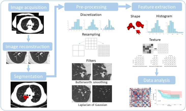
Radiomics has become a popular image analysis method in the last few years. Its key hypothesis is that medical images harbor biological, prognostic and predictive information that is not revealed upon visual inspection. In contrast to previous work with a priori defined imaging biomarkers, radiomics instead calculates image features at scale and uses statistical methods to identify those most strongly associated to outcome. This builds on years of research into computer aided diagnosis and pattern recognition. While the potential of radiomics to aid personalized medicine is widely recognized, several technical limitations exist which hinder biomarker translation. Aspects of the radiomic workflow lack repeatability or reproducibility under particular circumstances, which is a key requirement for the translation of imaging biomarkers into clinical practice. One of the most commonly studied uses of radiomics is for personalized medicine applications in Non-Small Cell Lung Cancer (NSCLC). In this review, we summarize reported methodological limitations in CT based radiomic analyses together with suggested solutions. We then evaluate the current NSCLC radiomics literature to assess the risk associated with accepting the published conclusions with respect to these limitations. We review different complementary scoring systems and initiatives that can be used to critically appraise data from radiomics studies. Wider awareness should improve the quality of ongoing and future radiomics studies and advance their potential as clinically relevant biomarkers for personalized medicine in patients with NSCLC.
近年来,放射组学已成为一种流行的医学图像分析方法。其关键假设是,医学图像中蕴藏着通过肉眼观察无法揭示的生物学、预后和预测信息。与先前使用先验定义的成像生物标志物的工作不同,放射组学可以大规模地计算图像特征,并使用统计方法来识别与结果最相关的特征。这是基于多年来对计算机辅助诊断和模式识别的研究。尽管放射组学在辅助个性化医疗方面的潜力已被广泛认可,但存在一些技术限制,阻碍了生物标志物的转化。在特定情况下,放射组学工作流程的某些方面缺乏可重复性或可再现性,这是将成像生物标志物转化为临床实践的关键要求。放射组学最常被研究的用途之一是在非小细胞肺癌(NSCLC)的个性化医疗应用中。在这篇综述中,我们总结了 CT 基放射组学分析中报道的方法学限制以及提出的解决方案。然后,我们评估了当前 NSCLC 放射组学文献,以评估接受这些限制下发表的结论所带来的风险。我们回顾了不同的补充评分系统和计划,可用于批判性地评估放射组学研究的数据。更广泛的认识应该提高正在进行和未来的放射组学研究的质量,并推进其作为 NSCLC 患者个性化医疗中具有临床相关性的生物标志物的潜力。