Huynh Elizabeth, Coroller Thibaud P, Narayan Vivek, Agrawal Vishesh, Romano John, Franco Idalid, Parmar Chintan, Hou Ying, Mak Raymond H, Aerts Hugo J W L
Department of Radiation Oncology, Dana-Farber Cancer Institute, Brigham and Women's Hospital, Harvard Medical School, Boston, United States of America.
Department of Radiology, Dana-Farber Cancer Institute, Brigham and Women's Hospital, Harvard Medical School, Boston, United States of America.
PLoS One. 2017 Jan 3;12(1):e0169172. doi: 10.1371/journal.pone.0169172. eCollection 2017.
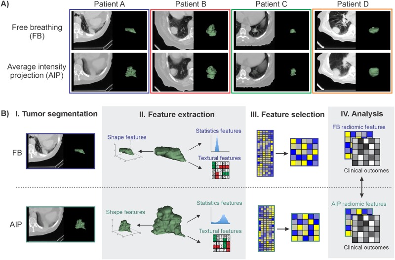
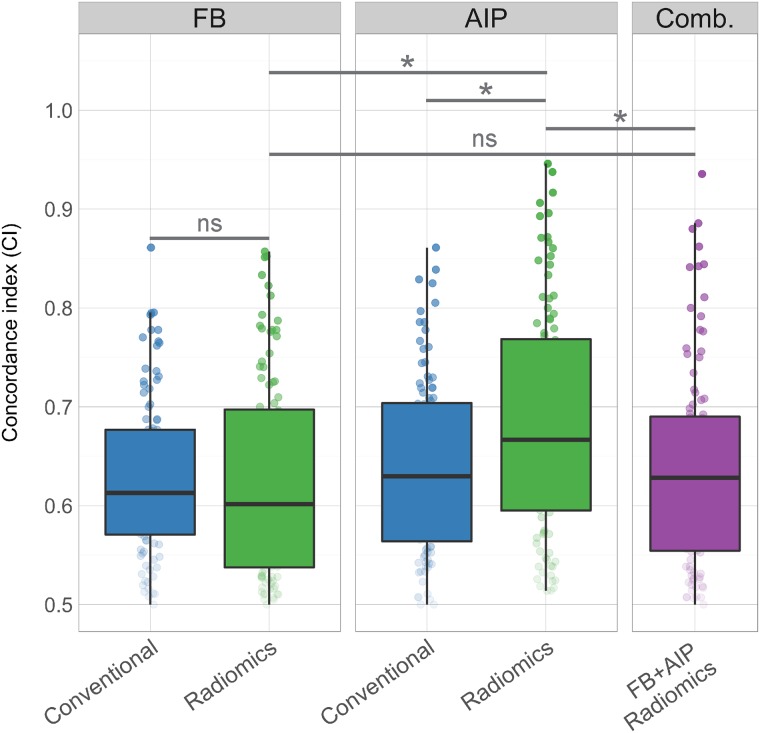
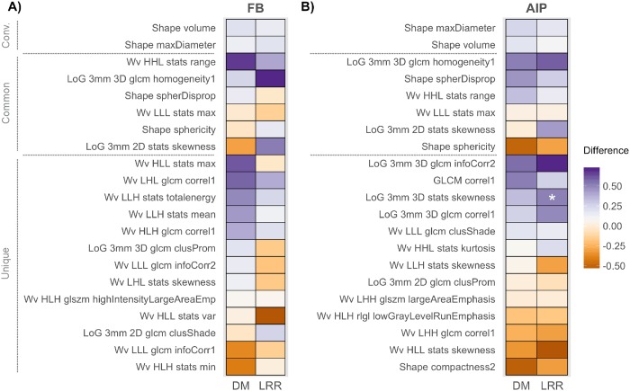
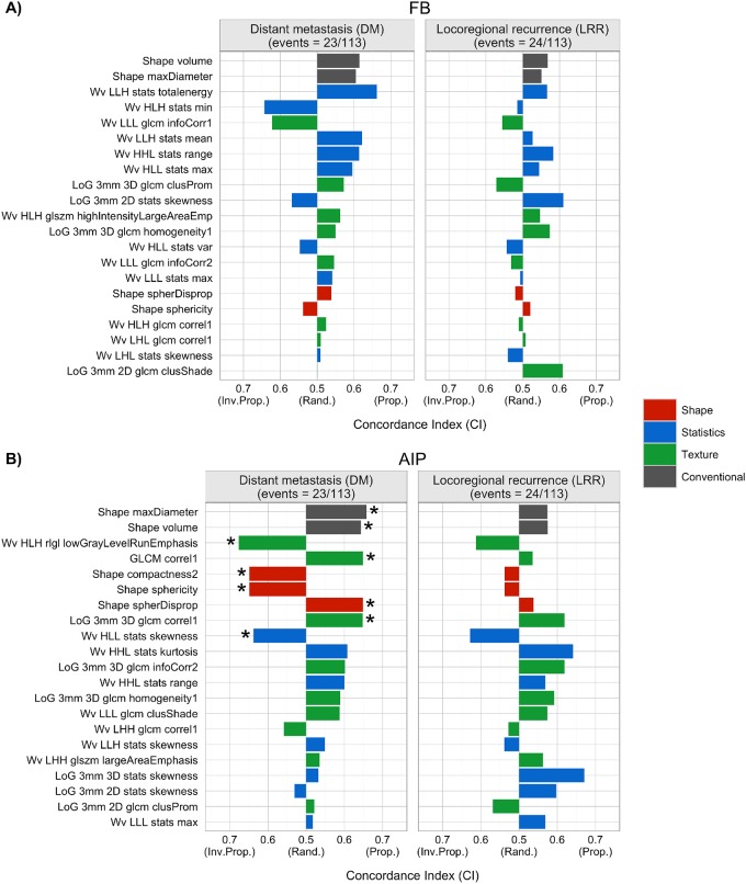
Radiomics aims to quantitatively capture the complex tumor phenotype contained in medical images to associate them with clinical outcomes. This study investigates the impact of different types of computed tomography (CT) images on the prognostic performance of radiomic features for disease recurrence in early stage non-small cell lung cancer (NSCLC) patients treated with stereotactic body radiation therapy (SBRT). 112 early stage NSCLC patients treated with SBRT that had static free breathing (FB) and average intensity projection (AIP) images were analyzed. Nineteen radiomic features were selected from each image type (FB or AIP) for analysis based on stability and variance. The selected FB and AIP radiomic feature sets had 6 common radiomic features between both image types and 13 unique features. The prognostic performances of the features for distant metastasis (DM) and locoregional recurrence (LRR) were evaluated using the concordance index (CI) and compared with two conventional features (tumor volume and maximum diameter). P-values were corrected for multiple testing using the false discovery rate procedure. None of the FB radiomic features were associated with DM, however, seven AIP radiomic features, that described tumor shape and heterogeneity, were (CI range: 0.638-0.676). Conventional features from FB images were not associated with DM, however, AIP conventional features were (CI range: 0.643-0.658). Radiomic and conventional multivariate models were compared between FB and AIP images using cross validation. The differences between the models were assessed using a permutation test. AIP radiomic multivariate models (median CI = 0.667) outperformed all other models (median CI range: 0.601-0.630) in predicting DM. None of the imaging features were prognostic of LRR. Therefore, image type impacts the performance of radiomic models in their association with disease recurrence. AIP images contained more information than FB images that were associated with disease recurrence in early stage NSCLC patients treated with SBRT, which suggests that AIP images may potentially be more optimal for the development of an imaging biomarker.
放射组学旨在定量获取医学图像中包含的复杂肿瘤表型,以便将其与临床结果相关联。本研究调查了不同类型的计算机断层扫描(CT)图像对接受立体定向体部放射治疗(SBRT)的早期非小细胞肺癌(NSCLC)患者疾病复发的放射组学特征预后性能的影响。分析了112例接受SBRT治疗且有静态自由呼吸(FB)和平均强度投影(AIP)图像的早期NSCLC患者。基于稳定性和方差,从每种图像类型(FB或AIP)中选择了19个放射组学特征进行分析。所选的FB和AIP放射组学特征集在两种图像类型之间有6个共同的放射组学特征和13个独特特征。使用一致性指数(CI)评估这些特征对远处转移(DM)和局部区域复发(LRR)的预后性能,并与两个传统特征(肿瘤体积和最大直径)进行比较。使用错误发现率程序对多重检验的P值进行校正。没有一个FB放射组学特征与DM相关,然而,七个描述肿瘤形状和异质性的AIP放射组学特征与DM相关(CI范围:0.638 - 0.676)。FB图像的传统特征与DM无关,然而,AIP传统特征与DM相关(CI范围:0.643 - 0.658)。使用交叉验证比较了FB和AIP图像之间的放射组学和传统多变量模型。使用置换检验评估模型之间的差异。在预测DM方面,AIP放射组学多变量模型(中位数CI = 0.667)优于所有其他模型(中位数CI范围:0.601 - 0.630)。没有一个成像特征对LRR具有预后价值。因此,图像类型会影响放射组学模型与疾病复发关联的性能。在接受SBRT治疗的早期NSCLC患者中,AIP图像比FB图像包含更多与疾病复发相关的信息,这表明AIP图像可能对成像生物标志物的开发更具潜力。