Department of Biochemistry and Molecular Biology, Nippon Medical School, Tokyo, Japan.
Department of Drug Discovery Research, Teika Pharmaceutical Co., Ltd., Toyama, Japan.
Mol Vis. 2020 Jun 3;26:409-422. eCollection 2020.
Glaucoma is a group of chronic optic neuropathies characterized by the degeneration of retinal ganglion cells (RGCs) and their axons, and they ultimately cause blindness. Because neuroprotection using neurotrophic factors against RGC loss has been proven a beneficial strategy, extensive attempts have been made to perform gene transfer of neurotrophic proteins. This study used the inner retinal injury mouse model to evaluate the neuroprotective effect of tyrosine triple mutated and self-complementary adeno-associated virus (AAV) encoding brain-derived neurotrophic factor (BDNF; tm-scAAV2-BDNF).
C57BL/6J mice were intravitreally injected with 1 μl of tm-scAAV2-BDNF and its control AAV at a titer of 6.6 E+13 genome copies/ml. Three weeks later, 1 μl of 2 mM -methyl-D-aspartate (NMDA) was administered in the same way as the viral injection. Six days after the NMDA injection, we assessed the dark-adapted electroretinography (ERG). Mice were sacrificed at one week after the NMDA injection, followed by RNA quantification, protein detection, and histopathological analysis.
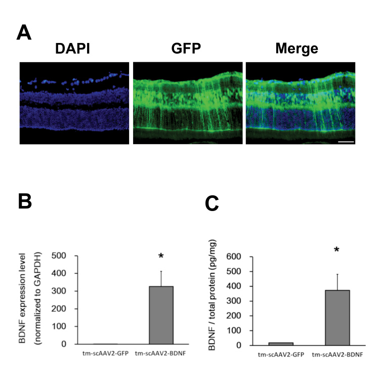
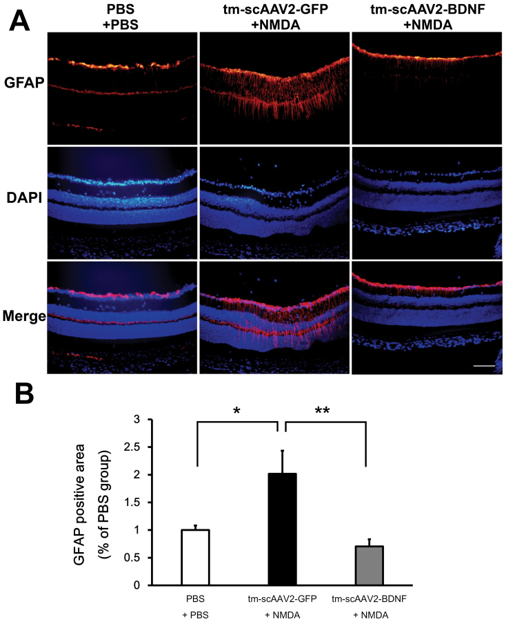
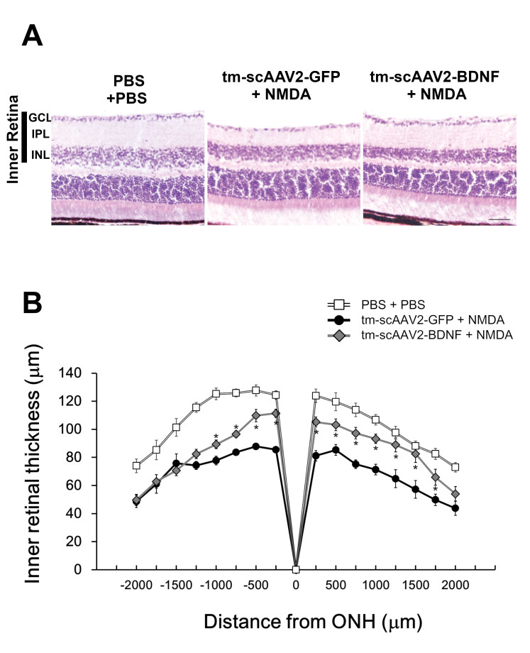
The RNA expression of BDNF in retinas treated with tm-scAAV2-BDNF was about 300-fold higher than that of its control AAV. Meanwhile, the expression of recombinant BDNF protein increased in retinas treated with tm-scAAV2-BDNF. In addition, histological analysis revealed that tm-scAAV2-BDNF prevented thinning of the inner retina. Furthermore, b-wave amplitudes of the tm-scAAV2-BDNF group were significantly higher than those of the control vector group. Histopathological and electrophysiological evaluations showed that tm-scAAV2-BDNF treatment offered significant protection against NMDA toxicity.
Results showed that tm-scAAV2-BDNF-treated retinas were resistant to NMDA injury, while retinas treated with the control AAV exhibited histopathological and functional changes after the administration of NMDA. These results suggest that tm-scAAV2-BDNF is potentially effective against inner retinal injury, including normal tension glaucoma.
青光眼是一组以视网膜神经节细胞(RGC)及其轴突变性为特征的慢性视神经病变,最终导致失明。由于神经营养因子对 RGC 丢失的神经保护作用已被证明是一种有益的策略,因此已经进行了广泛的尝试来进行神经营养蛋白的基因转移。本研究使用内视网膜损伤小鼠模型来评估酪氨酸三突变和自互补腺相关病毒(AAV)编码脑源性神经营养因子(BDNF;tm-scAAV2-BDNF)的神经保护作用。
将 1 μl 的 tm-scAAV2-BDNF 和其对照 AAV 以 6.6 E+13 基因组拷贝/ml 的滴度注入 C57BL/6J 小鼠的玻璃体腔。3 周后,以与病毒注射相同的方式向玻璃体腔内注射 1 μl 2 mM -甲基-D-天冬氨酸(NMDA)。NMDA 注射后 6 天,我们评估暗适应视网膜电图(ERG)。NMDA 注射后 1 周处死小鼠,然后进行 RNA 定量、蛋白质检测和组织病理学分析。
用 tm-scAAV2-BDNF 处理的视网膜中 BDNF 的 RNA 表达比其对照 AAV 高约 300 倍。同时,用 tm-scAAV2-BDNF 处理的视网膜中重组 BDNF 蛋白的表达增加。此外,组织学分析表明,tm-scAAV2-BDNF 可防止内视网膜变薄。此外,tm-scAAV2-BDNF 组的 b 波幅度明显高于对照载体组。组织病理学和电生理学评估表明,tm-scAAV2-BDNF 治疗对 NMDA 毒性具有显著的保护作用。
结果表明,用 tm-scAAV2-BDNF 处理的视网膜对 NMDA 损伤具有抗性,而用对照 AAV 处理的视网膜在给予 NMDA 后表现出组织病理学和功能变化。这些结果表明,tm-scAAV2-BDNF 对内视网膜损伤具有潜在的治疗作用,包括正常眼压性青光眼。