Department of Forensic Medicine and Toxicology, Faculty of Veterinary Medicine, University of Sadat City, Sadat City, Egypt.
Department of Biology, Faculty of Sciences and Arts-Khulais, University of Jeddah, Jeddah, Saudi Arabia.
Int J Nanomedicine. 2020 May 28;15:3827-3842. doi: 10.2147/IJN.S241922. eCollection 2020.
Copper oxide nanoparticles (CuO-NPs) are widely used as feed additives for livestock and poultry and implicated in many biomedical applications; however, overload of copper NPs induces various toxicological changes and dysfunction of animal's organs. Thus, this study was designed to evaluate the comparative toxicological effects of biologically and chemically synthesized CuO-NPs on mice.
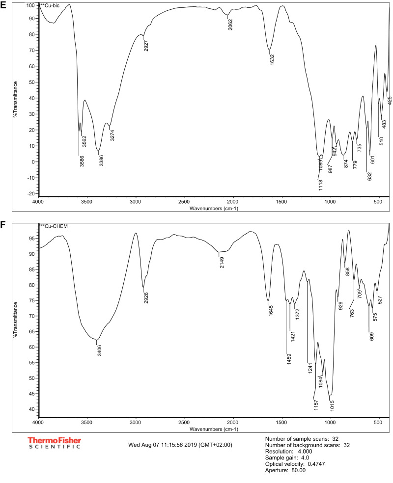
Transmission electron microscopy (TEM), X-ray diffraction (XRD) and Fourier-transform infrared spectroscopy (FT-IR) were used to characterize the sizes, shapes and functional groups of CuO-NPs. Forty-five mice were randomly allocated into three groups. Control group received distilled water. The second group was administered a single dose of biologically synthesized CuO-NPs (500 mg/kg bw) orally. The third group was administered a single dose of chemically synthesized CuO-NPs (500 mg/kg bw) orally.
TEM revealed that biologically synthesized NPs were spherical in shape, whereas chemically synthesized NPs were spherical or elongated in shape. XRD showed that the size of biologically synthesized NPs ranged from 4.14 to 12.82 nm and that of chemically synthesized NPs ranged from 4.06 to 26.82 nm. FT-IR spectroscopy indicated that the peaks appeared between 779 cm and 425 cm in biologically synthesized NPs and between 858 cm and 524 cm in chemically synthesized NPs were for Cu-O nanostructure. Four mice died due to administration of biologically synthesized CuO-NPs. Both biologically and chemically synthesized CuO-NPs induced leukocytosis, elevated serum activities of alanine aminotransferase and aspartate aminotransferase and serum levels of urea and creatinine and increased P53 mRNA and caspase-3 protein expressions in hepatic tissues. Moreover, CuO-NPs induced degenerative and necrotized changes in hepatic, renal and splenic tissues. Biochemical, apoptotic and pathological changes were more serious in mice administered with biologically synthesized CuO-NPs.
This study indicated that a high dose of biologically and chemically synthesized CuO-NPs induced adverse effects on hepatic, renal and splenic tissues. At the same dose level, the biologically synthesized CuO-NPs evoked more potent toxic effects than the chemically synthesized CuO-NPs.
氧化铜纳米粒子(CuO-NPs)被广泛用作饲料添加剂用于家畜和家禽,并被应用于许多生物医学领域;然而,铜纳米粒子的过载会引起动物器官的各种毒性变化和功能障碍。因此,本研究旨在评估生物合成和化学合成的 CuO-NPs 对小鼠的比较毒理学效应。
透射电子显微镜(TEM)、X 射线衍射(XRD)和傅里叶变换红外光谱(FT-IR)用于表征 CuO-NPs 的尺寸、形状和官能团。45 只小鼠被随机分为三组。对照组给予蒸馏水。第二组小鼠经口给予生物合成的 CuO-NPs(500mg/kg bw)单剂量。第三组小鼠经口给予化学合成的 CuO-NPs(500mg/kg bw)单剂量。
TEM 显示,生物合成的 NPs 呈球形,而化学合成的 NPs 呈球形或拉长形。XRD 表明,生物合成的 NPs 尺寸范围为 4.14-12.82nm,化学合成的 NPs 尺寸范围为 4.06-26.82nm。FT-IR 光谱表明,生物合成的 NPs 中在 779cm 至 425cm 之间出现的峰和化学合成的 NPs 中在 858cm 至 524cm 之间出现的峰为 Cu-O 纳米结构。由于给予生物合成的 CuO-NPs,有 4 只小鼠死亡。生物合成和化学合成的 CuO-NPs 均诱导白细胞增多、血清丙氨酸氨基转移酶和天冬氨酸氨基转移酶活性升高、血清尿素和肌酐水平升高以及肝组织 P53mRNA 和 caspase-3 蛋白表达增加。此外,CuO-NPs 诱导肝、肾和脾组织的退行性和坏死性变化。给予生物合成的 CuO-NPs 的小鼠的生化、凋亡和病理变化更为严重。
本研究表明,高剂量的生物合成和化学合成的 CuO-NPs 对肝、肾和脾组织产生不良影响。在相同剂量水平下,生物合成的 CuO-NPs 比化学合成的 CuO-NPs 产生更强的毒性作用。