Vavrdová Tereza, Křenek Pavel, Ovečka Miroslav, Šamajová Olga, Floková Pavlína, Illešová Petra, Šnaurová Renáta, Šamaj Jozef, Komis George
Department of Cell Biology, Centre of the Region Haná for Biotechnological and Agricultural Research, Faculty of Science, Palacký University Olomouc, Olomouc, Czechia.
Front Plant Sci. 2020 Jun 5;11:693. doi: 10.3389/fpls.2020.00693. eCollection 2020.
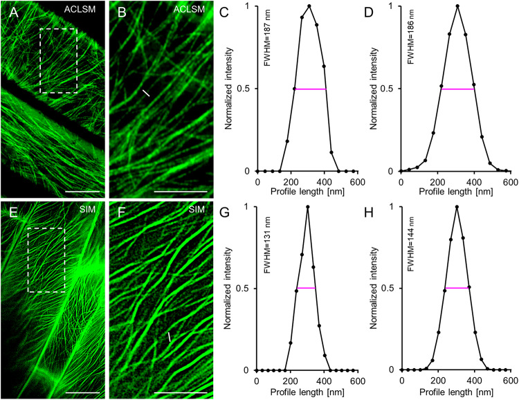
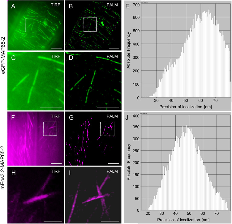
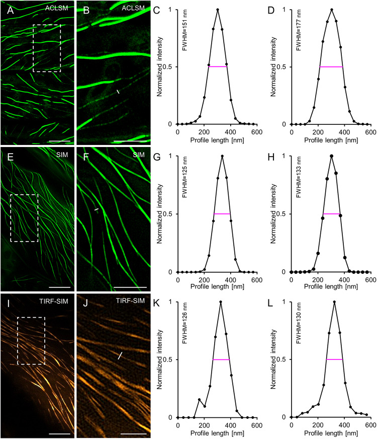
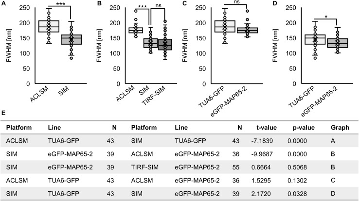
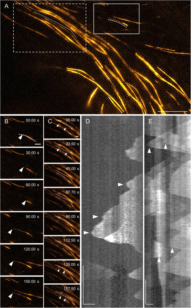
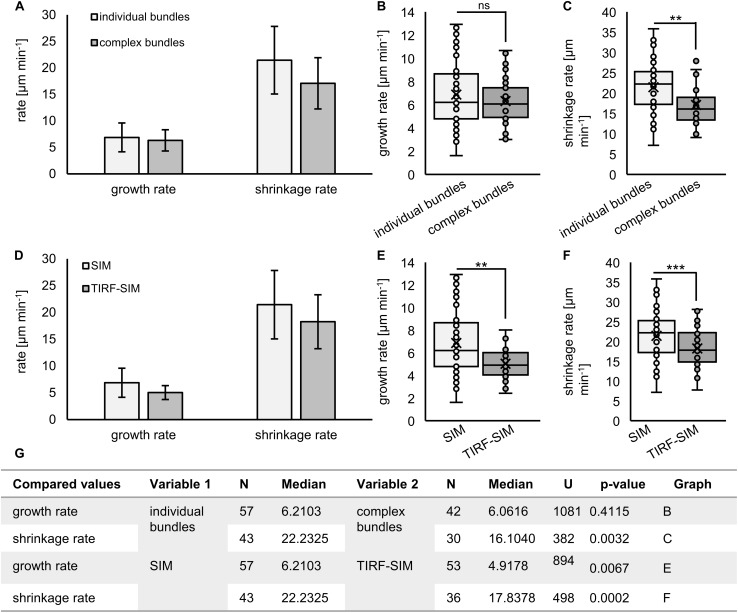
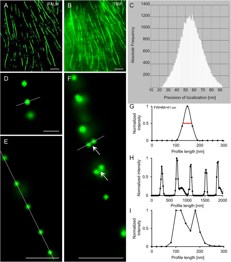
Microtubule bundling is an essential mechanism underlying the biased organization of interphase and mitotic microtubular systems of eukaryotes in ordered arrays. Microtubule bundle formation can be exemplified in plants, where the formation of parallel microtubule systems in the cell cortex or the spindle midzone is largely owing to the microtubule crosslinking activity of a family of microtubule associated proteins, designated as MAP65s. Among the nine members of this family in , MAP65-1 and MAP65-2 are ubiquitous and functionally redundant. Crosslinked microtubules can form high-order arrays, which are difficult to track using widefield or confocal laser scanning microscopy approaches. Here, we followed spatiotemporal patterns of MAP65-2 localization in hypocotyl cells of Arabidopsis stably expressing fluorescent protein fusions of MAP65-2 and tubulin. To circumvent imaging difficulties arising from the density of cortical microtubule bundles, we use different superresolution approaches including Airyscan confocal laser scanning microscopy (ACLSM), structured illumination microscopy (SIM), total internal reflection SIM (TIRF-SIM), and photoactivation localization microscopy (PALM). We provide insights into spatiotemporal relations between microtubules and MAP65-2 crossbridges by combining SIM and ACLSM. We obtain further details on MAP65-2 distribution by single molecule localization microscopy (SMLM) imaging of either mEos3.2-MAP65-2 stochastic photoconversion, or eGFP-MAP65-2 stochastic emission fluctuations under specific illumination conditions. Time-dependent dynamics of MAP65-2 were tracked at variable time resolution using SIM, TIRF-SIM, and ACLSM and post-acquisition kymograph analysis. ACLSM imaging further allowed to track end-wise dynamics of microtubules labeled with TUA6-GFP and to correlate them with concomitant fluctuations of MAP65-2 tagged with tagRFP. All different microscopy modules examined herein are accompanied by restrictions in either the spatial resolution achieved, or in the frame rates of image acquisition. PALM imaging is compromised by speed of acquisition. This limitation was partially compensated by exploiting emission fluctuations of eGFP which allowed much higher photon counts at substantially smaller time series compared to mEos3.2. SIM, TIRF-SIM, and ACLSM were the methods of choice to follow the dynamics of MAP65-2 in bundles of different complexity. Conclusively, the combination of different superresolution methods allowed for inferences on the distribution and dynamics of MAP65-2 within microtubule bundles of living cells.
微管成束是真核生物间期和有丝分裂微管系统在有序阵列中偏向性组织的一种基本机制。微管束的形成在植物中可以得到例证,在植物中,细胞皮层或纺锤体中区平行微管系统的形成很大程度上归因于一类微管相关蛋白(称为MAP65s)的微管交联活性。在该家族的九个成员中,MAP65-1和MAP65-2普遍存在且功能冗余。交联的微管可以形成高阶阵列,使用宽场或共聚焦激光扫描显微镜方法很难追踪这些阵列。在这里,我们追踪了稳定表达MAP65-2和微管蛋白荧光蛋白融合体的拟南芥下胚轴细胞中MAP65-2定位的时空模式。为了规避因皮层微管束密度而产生的成像困难,我们使用了不同的超分辨率方法,包括Airyscan共聚焦激光扫描显微镜(ACLSM)、结构光照显微镜(SIM)、全内反射SIM(TIRF-SIM)和光激活定位显微镜(PALM)。我们通过结合SIM和ACLSM,深入了解了微管与MAP65-2交联桥之间的时空关系。我们通过对mEos3.2-MAP65-2随机光转化或在特定光照条件下eGFP-MAP65-2随机发射波动进行单分子定位显微镜(SMLM)成像,获得了关于MAP65-2分布的更多细节。使用SIM、TIRF-SIM和ACLSM以及采集后制作的动态曲线图分析,以可变的时间分辨率追踪MAP65-2的时间依赖性动力学。ACLSM成像还可以追踪用TUA6-GFP标记的微管的末端动力学,并将它们与用tagRFP标记的MAP65-2的伴随波动相关联。本文研究的所有不同显微镜模块在实现的空间分辨率或图像采集帧率方面都存在限制。PALM成像受采集速度的影响。通过利用eGFP的发射波动部分弥补了这一限制,与mEos3.2相比,在时间序列小得多的情况下,eGFP允许更高的光子计数。SIM、TIRF-SIM和ACLSM是追踪不同复杂程度微管束中MAP65-2动力学的首选方法。总之,不同超分辨率方法的结合使得能够推断活细胞微管束中MAP65-2的分布和动力学。