• 文献检索
  • 文档翻译
  • 深度研究
  • 学术资讯
  • Suppr Zotero 插件Zotero 插件
  • 邀请有礼
  • 套餐&价格
  • 历史记录
应用&插件
Suppr Zotero 插件Zotero 插件浏览器插件Mac 客户端Windows 客户端微信小程序
定价
高级版会员购买积分包购买API积分包
服务
文献检索文档翻译深度研究API 文档MCP 服务
关于我们
关于 Suppr公司介绍联系我们用户协议隐私条款
关注我们

Suppr 超能文献

核心技术专利:CN118964589B侵权必究
粤ICP备2023148730 号-1Suppr @ 2026

文献检索

告别复杂PubMed语法,用中文像聊天一样搜索,搜遍4000万医学文献。AI智能推荐,让科研检索更轻松。

立即免费搜索

文件翻译

保留排版,准确专业,支持PDF/Word/PPT等文件格式,支持 12+语言互译。

免费翻译文档

深度研究

AI帮你快速写综述,25分钟生成高质量综述,智能提取关键信息,辅助科研写作。

立即免费体验

原子力显微镜和体内磁共振弹性成像研究小鼠海马亚区的力学性能。

Mechanical properties of murine hippocampal subregions investigated by atomic force microscopy and in vivo magnetic resonance elastography.

机构信息

Department of Radiology, Charité-Universitätsmedizin Berlin, Corporate Member of Freie Universität Berlin and Humboldt-Universität zu Berlin, Charitéplatz 1, 10117, Berlin, Germany.

Institute of Anatomy, University of Leipzig, Leipzig, Germany.

出版信息

Sci Rep. 2022 Oct 6;12(1):16723. doi: 10.1038/s41598-022-21105-7.

DOI:10.1038/s41598-022-21105-7
PMID:36202964
原文链接:https://pmc.ncbi.nlm.nih.gov/articles/PMC9537158/
Abstract

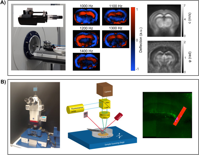
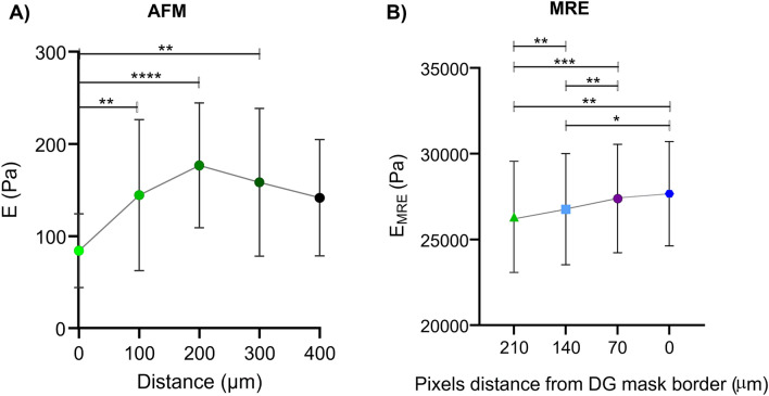
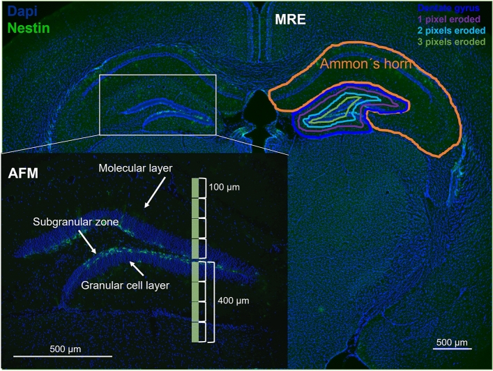
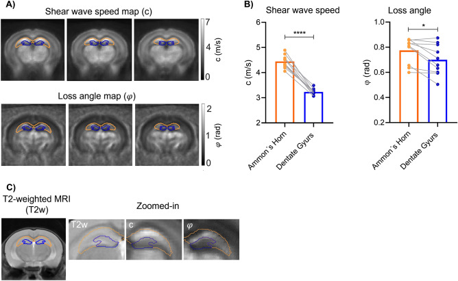
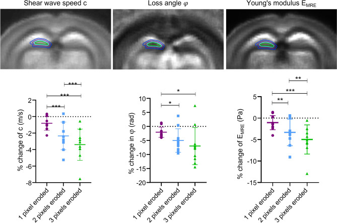
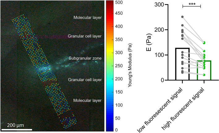
The hippocampus is a very heterogeneous brain structure with different mechanical properties reflecting its functional variety. In particular, adult neurogenesis in rodent hippocampus has been associated with specific viscoelastic properties in vivo and ex vivo. Here, we study the microscopic mechanical properties of hippocampal subregions using ex vivo atomic force microscopy (AFM) in correlation with the expression of GFP in presence of the nestin promoter, providing a marker of neurogenic activity. We further use magnetic resonance elastography (MRE) to investigate whether in vivo mechanical properties reveal similar spatial patterns, however, on a much coarser scale. AFM showed that tissue stiffness increases with increasing distance from the subgranular zone (p = 0.0069), and that stiffness is 39% lower in GFP than non-GFP regions (p = 0.0004). Consistently, MRE showed that dentate gyrus is, on average, softer than Ammon´s horn (shear wave speed = 3.2 ± 0.2 m/s versus 4.4 ± 0.3 m/s, p = 0.01) with another 3.4% decrease towards the subgranular zone (p = 0.0001). The marked reduction in stiffness measured by AFM in areas of high neurogenic activity is consistent with softer MRE values, indicating the sensitivity of macroscopic mechanical properties in vivo to micromechanical structures as formed by the neurogenic niche of the hippocampus.

摘要

海马体是一个非常异质的脑结构,具有不同的力学特性,反映了其功能的多样性。特别是,啮齿动物海马体中的成年神经发生与体内和体外的特定粘弹性特性有关。在这里,我们使用体外原子力显微镜(AFM)研究海马体亚区的微观力学特性,并与巢蛋白启动子存在下 GFP 的表达相关联,提供神经发生活性的标志物。我们进一步使用磁共振弹性成像(MRE)来研究体内力学特性是否揭示出类似的空间模式,然而,其在更粗糙的尺度上。AFM 显示,组织刚度随距颗粒下层(SGZ)距离的增加而增加(p=0.0069),并且 GFP 区域的刚度比非 GFP 区域低 39%(p=0.0004)。一致地,MRE 显示出齿状回的平均硬度低于 Ammon´s horn(剪切波速度=3.2±0.2 m/s 与 4.4±0.3 m/s,p=0.01),而向 SGZ 方向则降低了 3.4%(p=0.0001)。在具有高神经发生活性的区域中,AFM 测量的刚度明显降低与 MRE 值变软一致,表明体内宏观力学特性对海马体神经发生龛形成的微结构具有敏感性。

https://cdn.ncbi.nlm.nih.gov/pmc/blobs/75fc/9537158/86c2405d0f01/41598_2022_21105_Fig6_HTML.jpg
https://cdn.ncbi.nlm.nih.gov/pmc/blobs/75fc/9537158/cc49994602c3/41598_2022_21105_Fig1_HTML.jpg
https://cdn.ncbi.nlm.nih.gov/pmc/blobs/75fc/9537158/3573a4c20f1e/41598_2022_21105_Fig2_HTML.jpg
https://cdn.ncbi.nlm.nih.gov/pmc/blobs/75fc/9537158/c11bc86baf19/41598_2022_21105_Fig3_HTML.jpg
https://cdn.ncbi.nlm.nih.gov/pmc/blobs/75fc/9537158/63bcb9564247/41598_2022_21105_Fig4_HTML.jpg
https://cdn.ncbi.nlm.nih.gov/pmc/blobs/75fc/9537158/d3e4dcf4bf03/41598_2022_21105_Fig5_HTML.jpg
https://cdn.ncbi.nlm.nih.gov/pmc/blobs/75fc/9537158/86c2405d0f01/41598_2022_21105_Fig6_HTML.jpg

相似文献

1
Mechanical properties of murine hippocampal subregions investigated by atomic force microscopy and in vivo magnetic resonance elastography.原子力显微镜和体内磁共振弹性成像研究小鼠海马亚区的力学性能。
Sci Rep. 2022 Oct 6;12(1):16723. doi: 10.1038/s41598-022-21105-7.
2
Mechanical behavior of the hippocampus and corpus callosum: An attempt to reconcile ex vivo with in vivo and micro with macro properties.海马体和胼胝体的力学行为:尝试协调离体与在体以及微观与宏观特性。
J Mech Behav Biomed Mater. 2023 Feb;138:105613. doi: 10.1016/j.jmbbm.2022.105613. Epub 2022 Dec 9.
3
A Shift in Tissue Stiffness During Hippocampal Maturation Correlates to the Pattern of Neurogenesis and Composition of the Extracellular Matrix.海马体成熟过程中组织硬度的变化与神经发生模式及细胞外基质组成相关。
Front Aging Neurosci. 2021 Jul 30;13:709620. doi: 10.3389/fnagi.2021.709620. eCollection 2021.
4
Mechanical heterogeneity of the rat hippocampus measured by atomic force microscope indentation.通过原子力显微镜压痕测量大鼠海马体的机械异质性。
J Neurotrauma. 2007 May;24(5):812-22. doi: 10.1089/neu.2006.0169.
5
Mechanical properties of porcine brain tissue in vivo and ex vivo estimated by MR elastography.通过磁共振弹性成像估计猪脑组织在体内和体外的力学特性。
J Biomech. 2018 Mar 1;69:10-18. doi: 10.1016/j.jbiomech.2018.01.016. Epub 2018 Jan 31.
6
Regional assessment of in vivo myocardial stiffness using 3D magnetic resonance elastography in a porcine model of myocardial infarction.利用 3D 磁共振弹性成像技术在猪心肌梗死模型中进行体内心肌硬度的区域性评估。
Magn Reson Med. 2018 Jan;79(1):361-369. doi: 10.1002/mrm.26695. Epub 2017 Apr 5.
7
Magnetic resonance elastography of the brain: A study of feasibility and reproducibility using an ergonomic pillow-like passive driver.脑部磁共振弹性成像:一项使用符合人体工程学的枕头状被动驱动器的可行性和可重复性研究。
Magn Reson Imaging. 2019 Jun;59:68-76. doi: 10.1016/j.mri.2019.03.009. Epub 2019 Mar 8.
8
Microscopic magnetic resonance elastography of traumatic brain injury model.外伤性脑损伤模型的微观磁共振弹性成像。
J Neurosci Methods. 2011 Oct 15;201(2):296-306. doi: 10.1016/j.jneumeth.2011.08.019. Epub 2011 Aug 17.
9
Measurement of anisotropic mechanical properties in porcine brain white matter ex vivo using magnetic resonance elastography.应用磁共振弹性成像技术对猪脑白质的各向异性力学特性进行离体测量。
J Mech Behav Biomed Mater. 2018 Mar;79:30-37. doi: 10.1016/j.jmbbm.2017.11.045. Epub 2017 Dec 9.
10
Magnetic resonance elastography in nonlinear viscoelastic materials under load.加载下非线性黏弹性材料的磁共振弹性成像。
Biomech Model Mechanobiol. 2019 Feb;18(1):111-135. doi: 10.1007/s10237-018-1072-1. Epub 2018 Aug 27.

引用本文的文献

1
Softness or Stiffness What Contributes to Cancer and Cancer Metastasis?柔软还是坚硬?是什么导致了癌症及癌症转移?
Cells. 2025 Apr 12;14(8):584. doi: 10.3390/cells14080584.
2
The Networking Brain: How Extracellular Matrix, Cellular Networks, and Vasculature Shape the In Vivo Mechanical Properties of the Brain.《联网大脑:细胞外基质、细胞网络和脉管系统如何塑造大脑的体内机械特性》
Adv Sci (Weinh). 2024 Aug;11(31):e2402338. doi: 10.1002/advs.202402338. Epub 2024 Jun 14.
3
"Patchiness" in mechanical stiffness across a tumor as an early-stage marker for malignancy.

本文引用的文献

1
Whole tissue and single cell mechanics are correlated in human brain tumors.在人脑肿瘤中,全组织力学和单细胞力学相互关联。
Soft Matter. 2021 Dec 8;17(47):10744-10752. doi: 10.1039/d1sm01291f.
2
Contribution of Tissue Inflammation and Blood-Brain Barrier Disruption to Brain Softening in a Mouse Model of Multiple Sclerosis.组织炎症和血脑屏障破坏对多发性硬化症小鼠模型脑软化的作用
Front Neurosci. 2021 Aug 23;15:701308. doi: 10.3389/fnins.2021.701308. eCollection 2021.
3
A Shift in Tissue Stiffness During Hippocampal Maturation Correlates to the Pattern of Neurogenesis and Composition of the Extracellular Matrix.
肿瘤机械硬度的“不均匀性”作为恶性肿瘤的早期标志物。
BMC Ecol Evol. 2024 Mar 14;24(1):33. doi: 10.1186/s12862-024-02221-6.
4
Photoacoustic viscoelasticity assessment of prefrontal cortex and cerebellum in normal and prenatal valproic acid-exposed rats.正常及产前暴露于丙戊酸的大鼠前额叶皮质和小脑的光声粘弹性评估
Photoacoustics. 2024 Jan 20;36:100590. doi: 10.1016/j.pacs.2024.100590. eCollection 2024 Apr.
5
Cortical matrix remodeling as a hallmark of relapsing-remitting neuroinflammation in MR elastography and quantitative MRI.皮质基质重塑作为磁共振弹性成像和定量磁共振中复发缓解性神经炎症的标志。
Acta Neuropathol. 2024 Jan 4;147(1):8. doi: 10.1007/s00401-023-02658-x.
6
Photoacoustic elasto-viscography and optical coherence microscopy for multi-parametric brain imaging.用于多参数脑成像的光声弹性黏滞成像和光学相干显微镜技术
Biomed Opt Express. 2023 Oct 6;14(11):5615-5628. doi: 10.1364/BOE.503847. eCollection 2023 Nov 1.
7
Combining atomic force microscopy and fluorescence-based techniques to explore mechanical properties of naive and ischemia-affected brain regions in mice.结合原子力显微镜和基于荧光的技术来探索小鼠未受影响和受缺血影响的脑区的机械性能。
Sci Rep. 2023 Aug 7;13(1):12774. doi: 10.1038/s41598-023-39277-1.
8
Hippocampus of the APP mouse model of Alzheimer's disease exhibits region-specific tissue softening concomitant with elevated astrogliosis.阿尔茨海默病APP小鼠模型的海马体表现出区域特异性组织软化,并伴有星形胶质细胞增生增加。
Front Aging Neurosci. 2023 Jul 20;15:1212212. doi: 10.3389/fnagi.2023.1212212. eCollection 2023.
海马体成熟过程中组织硬度的变化与神经发生模式及细胞外基质组成相关。
Front Aging Neurosci. 2021 Jul 30;13:709620. doi: 10.3389/fnagi.2021.709620. eCollection 2021.
4
Mechanical alterations of the hippocampus in the APP/PS1 Alzheimer's disease mouse model.APP/PS1 阿尔茨海默病小鼠模型中海马体的机械改变。
J Mech Behav Biomed Mater. 2021 Oct;122:104697. doi: 10.1016/j.jmbbm.2021.104697. Epub 2021 Jul 10.
5
Longitudinal study of sub-regional cerebral viscoelastic properties of 5XFAD Alzheimer's disease mice using multifrequency MR elastography.多频磁共振弹性成像研究 5XFAD 阿尔茨海默病小鼠脑区域性黏弹特性的纵向变化。
Magn Reson Med. 2021 Jul;86(1):405-414. doi: 10.1002/mrm.28709. Epub 2021 Feb 18.
6
Effect of Aging on the Viscoelastic Properties of Hippocampal Subfields Assessed with High-Resolution MR Elastography.衰老对通过高分辨率磁共振弹性成像评估的海马亚区粘弹性特性的影响。
Cereb Cortex. 2021 May 10;31(6):2799-2811. doi: 10.1093/cercor/bhaa388.
7
Superviscous properties of the in vivo brain at large scales.体内大脑在大尺度下的超粘性特性。
Acta Biomater. 2021 Feb;121:393-404. doi: 10.1016/j.actbio.2020.12.027. Epub 2020 Dec 14.
8
Viscoelastic mapping of mouse brain tissue: Relation to structure and age.小鼠脑组织的粘弹性映射:与结构和年龄的关系。
J Mech Behav Biomed Mater. 2021 Jan;113:104159. doi: 10.1016/j.jmbbm.2020.104159. Epub 2020 Oct 28.
9
Hippocampal stiffness in mesial temporal lobe epilepsy measured with MR elastography: Preliminary comparison with healthy participants.磁共振弹性成像测量内侧颞叶癫痫海马硬度:与健康参与者的初步比较。
Neuroimage Clin. 2020;27:102313. doi: 10.1016/j.nicl.2020.102313. Epub 2020 Jun 16.
10
Magnetic Resonance Elastography of Human Hippocampal Subfields: CA3-Dentate Gyrus Viscoelasticity Predicts Relational Memory Accuracy.磁共振弹性成像技术在人类海马亚区的应用:CA3-齿状回粘弹性预测关系记忆准确性。
J Cogn Neurosci. 2020 Sep;32(9):1704-1713. doi: 10.1162/jocn_a_01574. Epub 2020 May 7.