Department of Pediatrics, University of Arizona, Tucson, Arizona.
Department of Cellular and Molecular Medicine, University of Arizona, Tucson, Arizona.
Gastroenterology. 2020 Oct;159(4):1342-1356.e6. doi: 10.1053/j.gastro.2020.06.049. Epub 2020 Jun 23.
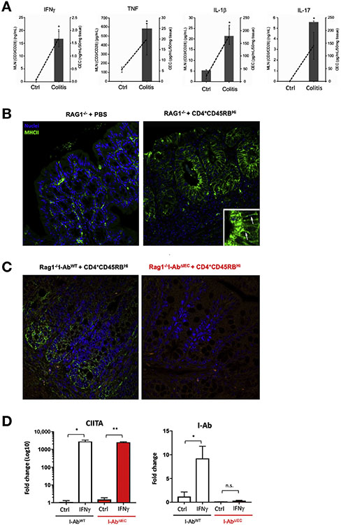
BACKGROUND & AIMS: Intestinal epithelial cells (IECs) provide a barrier that separates the mucosal immune system from the luminal microbiota. IECs constitutively express low levels of major histocompatibility complex (MHC) class II proteins, which are upregulated upon exposure to interferon gamma. We investigated the effects of deleting MHCII proteins specifically in mice with infectious, dextran sodium sulfate (DSS)-, and T-cell-induced colitis.
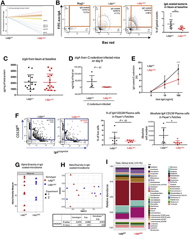
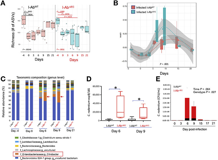
We disrupted the histocompatibility 2, class II antigen A, beta 1 gene (H2-Ab1) in IECs of C57BL/6 mice (I-Ab) or Rag1 mice (Rag1I-Ab); we used I-Ab mice as controls. Colitis was induced by administration of DSS, transfer of CD4CD45RB T cells, or infection with Citrobacter rodentium. Colon tissues were collected and analyzed by histology, immunofluorescence, xMAP, and reverse-transcription polymerase chain reaction and organoids were generated. Microbiota (total and immunoglobulin [Ig]A-coated) in intestinal samples were analyzed by16S amplicon profiling. IgACD138 plasma cells from Peyer's patches and lamina propria were analyzed by flow cytometry and IgA repertoire was determined by next-generation sequencing.
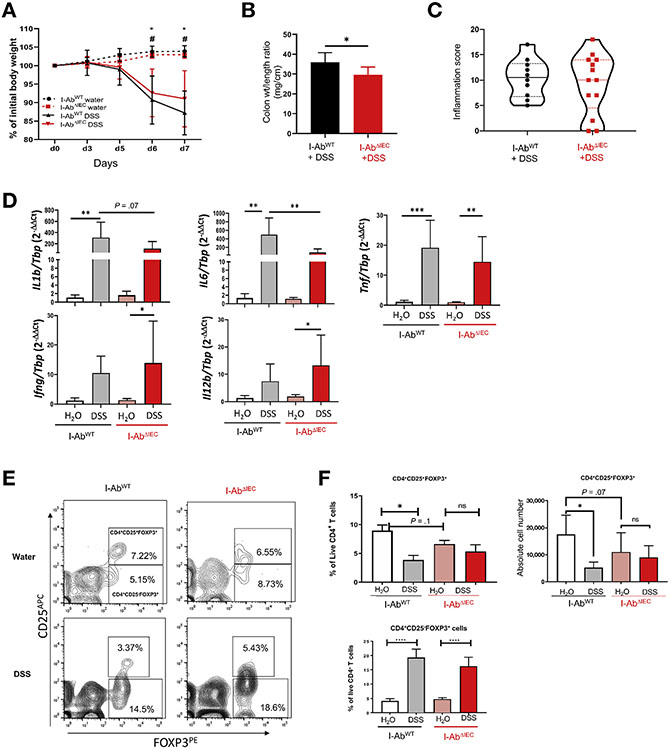
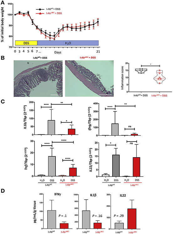
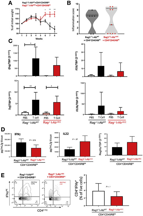
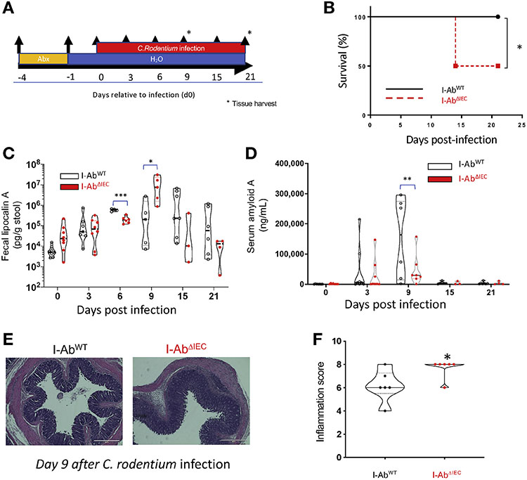
Mice with IEC-specific loss of MHCII (I-Ab mice) developed less severe DSS- or T-cell transfer-induced colitis than control mice. Intestinal tissues from I-Ab mice had a lower proportion of IgA-coated bacteria compared with control mice, and a reduced luminal concentration of secretory IgA (SIgA) following infection with C rodentium. There was no significant difference in the mucosal IgA repertoire of I-Ab vs control mice, but opsonization of cultured C rodentium by SIgA isolated from I-Ab mice was 50% lower than that of SIgA from mAb mice. Fifty percent of I-Ab mice died after infection with C rodentium, compared with none of the control mice. We observed a transient but significant expansion of the pathogen in the feces of I-Ab mice compared with I-Ab mice.
In mice with DSS or T-cell-induced colitis, loss of MHCII from IECs reduces but does not eliminate mucosal inflammation. However, in mice with C rodentium-induced colitis, loss of MHCII reduces bacterial clearance by decreasing binding of IgA to commensal and pathogenic bacteria.
肠上皮细胞(IECs)提供了一个屏障,将黏膜免疫系统与腔微生物群分开。IECs 持续低表达主要组织相容性复合体(MHC)II 类蛋白,这些蛋白在暴露于干扰素 γ 时会上调。我们研究了在具有传染性、葡聚糖硫酸钠(DSS)和 T 细胞诱导的结肠炎的小鼠中特异性缺失 MHCII 蛋白的影响。
我们在 C57BL/6 小鼠(I-Ab)或 Rag1 小鼠(Rag1I-Ab)的 IEC 中破坏组织相容性 2 类抗原 A、β1 基因(H2-Ab1);我们使用 I-Ab 小鼠作为对照。通过给予 DSS、转移 CD4+CD45RB T 细胞或感染柠檬酸杆菌来诱导结肠炎。收集结肠组织并进行组织学、免疫荧光、xMAP 和逆转录聚合酶链反应分析,并生成类器官。通过 16S 扩增子谱分析分析肠道样本中的微生物群(总微生物群和 IgA 包被的微生物群)。通过流式细胞术分析派尔集合淋巴结和固有层中的 IgACD138 浆细胞,并通过下一代测序确定 IgA 库。
与对照小鼠相比,具有 IEC 特异性 MHCII 缺失(I-Ab 小鼠)的小鼠发展出更严重的 DSS 或 T 细胞转移诱导的结肠炎。与对照小鼠相比,I-Ab 小鼠的肠道组织中 IgA 包被细菌的比例较低,感染柠檬酸杆菌后腔分泌型 IgA(SIgA)的浓度降低。I-Ab 与对照小鼠的黏膜 IgA 库没有显著差异,但从 I-Ab 小鼠分离的 SIgA 对培养的柠檬酸杆菌的调理作用比来自 mAb 小鼠的 SIgA 低 50%。50%的 I-Ab 感染柠檬酸杆菌的小鼠死亡,而对照小鼠无一死亡。与 I-Ab 小鼠相比,我们观察到 I-Ab 小鼠粪便中的病原体短暂但显著扩张。
在 DSS 或 T 细胞诱导的结肠炎小鼠中,IEC 中 MHCII 的缺失减少但不能消除黏膜炎症。然而,在柠檬酸杆菌诱导的结肠炎小鼠中,MHCII 的缺失通过减少 IgA 与共生菌和病原菌的结合来降低细菌清除率。