Division of Neurosurgery, Department of Neurology, Universidade de Sao Paulo, Sao Paulo, São Paulo, Brazil.
Department of Neuroradiology, Toronto Western Hospital, Toronto, Ontario, Canada
J Neurointerv Surg. 2021 Mar;13(3):272-277. doi: 10.1136/neurintsurg-2020-015990. Epub 2020 Jun 29.
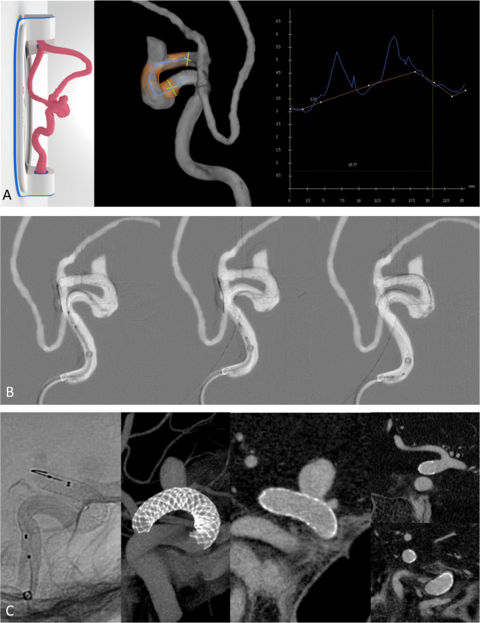
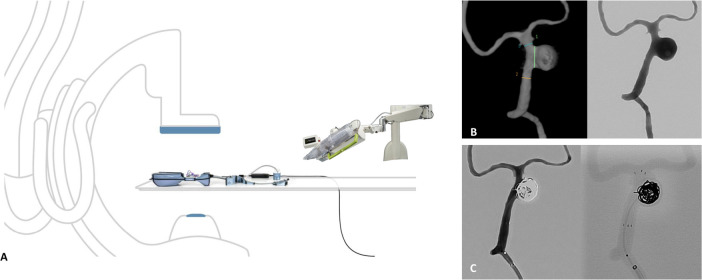
With the recent advent of advanced technologies in the field, treatment of neurovascular diseases using endovascular techniques is rapidly evolving. Here we describe our experience with pre-surgical simulation using the Biomodex EVIAS patient-specific 3D-printed models to plan aneurysm treatment using endovascular robotics and novel flow diverter devices.
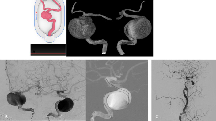
Pre-procedural rehearsals with 3D-printed patient-specific models of eight cases harboring brain aneurysms were performed before the first in-human experiences. To assess the reliability of the experimental model, the characteristics of the aneurysms were compared between the patient and 3D models. The rehearsals were used to define the patient treatment plan, including technique, device sizing, and operative working projections.
The study included eight patients with their respective EVIAS 3D aneurysm models. Pre-operative simulation was performed for the first in-human robotic-assisted neurovascular interventions (n=2) and new generation flow-diverter stents (n=6). Aneurysms were located in both the anterior (n=5) and posterior (n=3) circulation and were on average 11.0±6.5 mm in size. We found reliable reproduction of the aneurysm features and similar dimensions of the parent vessel anatomy between the 3D models and patient anatomy. Information learned from pre-surgical in vitro simulation are described in detail, including an improved patient treatment plan, which contributed to successful first in-world procedures with no intraprocedural complications.
Pre-procedural rehearsal using patient-specific 3D models provides precise procedure planning, which can potentially lead to greater operator confidence, decreased radiation dose and improvements in patient safety, particularly in first in-human experiences.
随着该领域先进技术的最新出现,使用血管内技术治疗神经血管疾病正在迅速发展。在这里,我们描述了使用 Biomodex EVIAS 患者特定的 3D 打印模型进行术前模拟的经验,以规划使用血管内机器人和新型血流导向装置治疗动脉瘤。
在首例人体试验之前,对 8 例颅内动脉瘤患者的 3D 打印患者特定模型进行了术前预演。为了评估实验模型的可靠性,将患者和 3D 模型之间的动脉瘤特征进行了比较。这些预演用于定义患者的治疗计划,包括技术、器械尺寸和手术工作投影。
该研究包括 8 例具有各自 EVIAS 3D 动脉瘤模型的患者。对首例机器人辅助神经血管介入治疗(n=2)和新一代血流导向支架(n=6)进行了术前模拟。动脉瘤位于前循环(n=5)和后循环(n=3),平均大小为 11.0±6.5mm。我们发现 3D 模型与患者解剖结构之间可以可靠地再现动脉瘤特征和相似的载瘤动脉解剖结构。详细描述了术前体外模拟中获得的信息,包括改进的患者治疗计划,这有助于成功完成首例人体试验,且无术中并发症。
使用患者特定的 3D 模型进行术前预演可提供精确的手术计划,这可能会提高操作人员的信心、降低辐射剂量并提高患者安全性,尤其是在首例人体试验中。