Institute of Fundamental Medicine and Biology, Kazan (Volga Region) Federal University, Kazan, Russia, 420008.
M.M. Shemyakin-Yu.A. Ovchinnikov Institute of Bioorganic Chemistry of the Russian Academy of Sciences, Moscow, Russia, 117997.
Sci Rep. 2020 Jul 1;10(1):10740. doi: 10.1038/s41598-020-67563-9.
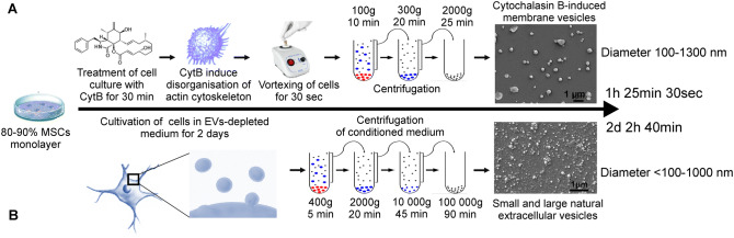
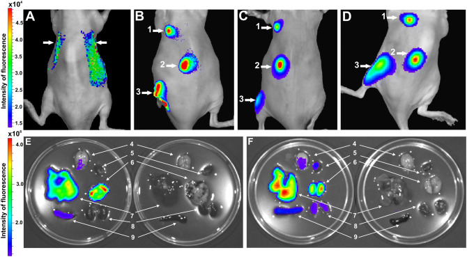
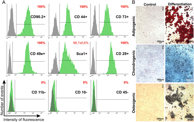
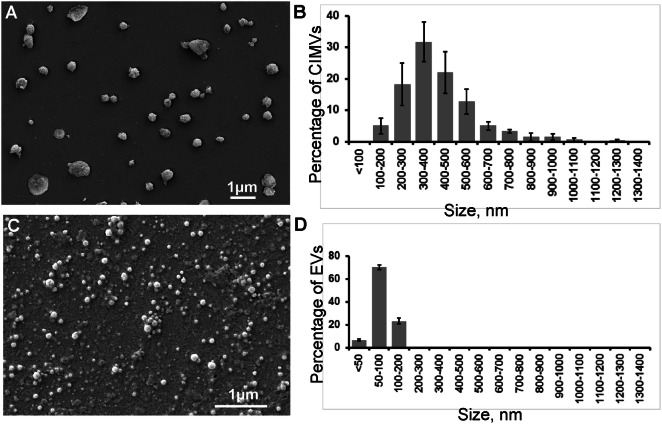
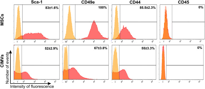
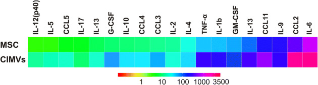
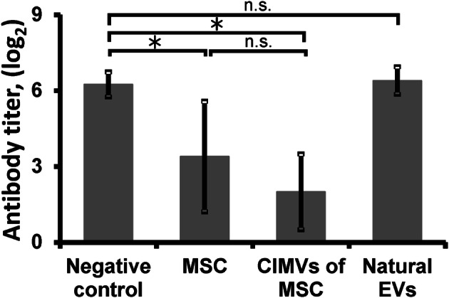
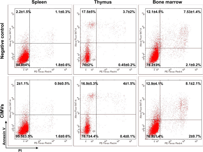
Extracellular vesicles derived from mesenchymal stem cells (MSCs) represent a novel approach for regenerative and immunosuppressive therapy. Recently, cytochalasin B-induced microvesicles (CIMVs) were shown to be effective drug delivery mediators. However, little is known about their immunological properties. We propose that the immunophenotype and molecular composition of these vesicles could contribute to the therapeutic efficacy of CIMVs. To address this issue, CIMVs were generated from murine MSC (CIMVs-MSCs) and their cytokine content and surface marker expression determined. For the first time, we show that CIMVs-MSCs retain parental MSCs phenotype (Sca-1, CD49e, CD44, CD45). Also, CIMVs-MSCs contained a cytokine repertoire reflective of the parental MSCs, including IL-1β, IL-2, IL-3, IL-4, IL-5, IL-6, IL-9, IL-10, IL-12(p40), IL-13, IL-17, CCL2, CCL3, CCL4, CCL5, CCL11, G-CSF, GM-CSF and TNF-α. Next, we evaluated the immune-modulating properties of CIMVs-MSCs in vivo using standard preclinical tests. MSCs and CIMVs-MSCs reduced serum levels of anti-sheep red blood cell antibody and have limited effects on neutrophil and peritoneal macrophage activity. We compared the immunomodulatory effect of MSCs, CIMVs and EVs. We observed no immunosuppression in mice pretreated with natural EVs, whereas MSCs and CIMVs-MSCs suppressed antibody production in vivo. Additionally, we have investigated the biodistribution of CIMVs-MSCs in vivo and demonstrated that CIMVs-MSCs localized in liver, lung, brain, heart, spleen and kidneys 48 h after intravenous injection and can be detected 14 days after subcutaneous and intramuscular injection. Collectively our data demonstrates immunomodulatory efficacy of CIMVs and supports their further preclinical testing as an effective therapeutic delivery modality.
间充质干细胞(MSCs)衍生的细胞外囊泡代表了一种用于再生和免疫抑制治疗的新方法。最近,细胞松弛素 B 诱导的微囊泡(CIMVs)已被证明是有效的药物传递介质。然而,对于它们的免疫学特性知之甚少。我们假设这些囊泡的免疫表型和分子组成可能有助于 CIMVs 的治疗效果。为了解决这个问题,我们从鼠 MSC 中生成了 CIMVs(CIMVs-MSCs),并确定了它们的细胞因子含量和表面标志物表达。我们首次表明,CIMVs-MSCs 保留了亲本 MSC 的表型(Sca-1、CD49e、CD44、CD45)。此外,CIMVs-MSCs 含有反映亲本 MSC 的细胞因子谱,包括 IL-1β、IL-2、IL-3、IL-4、IL-5、IL-6、IL-9、IL-10、IL-12(p40)、IL-13、IL-17、CCL2、CCL3、CCL4、CCL5、CCL11、G-CSF、GM-CSF 和 TNF-α。接下来,我们使用标准的临床前测试评估了 CIMVs-MSCs 在体内的免疫调节特性。MSCs 和 CIMVs-MSCs 降低了血清抗绵羊红细胞抗体的水平,对中性粒细胞和腹腔巨噬细胞的活性影响有限。我们比较了 MSCs、CIMVs 和 EVs 的免疫调节作用。我们观察到,用天然 EV 预处理的小鼠没有出现免疫抑制,而 MSCs 和 CIMVs-MSCs 则抑制了体内抗体的产生。此外,我们还研究了 CIMVs-MSCs 在体内的分布情况,并证明了 CIMVs-MSCs 在静脉注射后 48 小时定位在肝脏、肺、脑、心脏、脾脏和肾脏中,并且可以在皮下和肌肉内注射后 14 天检测到。总的来说,我们的数据证明了 CIMVs 的免疫调节功效,并支持进一步对其进行临床前测试,作为一种有效的治疗传递方式。