• 文献检索
  • 文档翻译
  • 深度研究
  • 学术资讯
  • Suppr Zotero 插件Zotero 插件
  • 邀请有礼
  • 套餐&价格
  • 历史记录
应用&插件
Suppr Zotero 插件Zotero 插件浏览器插件Mac 客户端Windows 客户端微信小程序
定价
高级版会员购买积分包购买API积分包
服务
文献检索文档翻译深度研究API 文档MCP 服务
关于我们
关于 Suppr公司介绍联系我们用户协议隐私条款
关注我们

Suppr 超能文献

核心技术专利:CN118964589B侵权必究
粤ICP备2023148730 号-1Suppr @ 2026

文献检索

告别复杂PubMed语法,用中文像聊天一样搜索,搜遍4000万医学文献。AI智能推荐,让科研检索更轻松。

立即免费搜索

文件翻译

保留排版,准确专业,支持PDF/Word/PPT等文件格式,支持 12+语言互译。

免费翻译文档

深度研究

AI帮你快速写综述,25分钟生成高质量综述,智能提取关键信息,辅助科研写作。

立即免费体验

表达Prx1的骨骼细胞及Prx1表达在骨折修复中的作用

Role of Prx1-expressing skeletal cells and Prx1-expression in fracture repair.

作者信息

Esposito Alessandra, Wang Lai, Li Tieshi, Miranda Mariana, Spagnoli Anna

机构信息

Department of Orthopaedic Surgery, Section of Molecular Medicine, Rush University Medical Center, Chicago, IL, USA.

Department of Internal Medicine, Division of Rheumatology, Rush University Medical Center, Chicago, IL, USA.

出版信息

Bone. 2020 Oct;139:115521. doi: 10.1016/j.bone.2020.115521. Epub 2020 Jul 3.

DOI:10.1016/j.bone.2020.115521
PMID:32629173
原文链接:https://pmc.ncbi.nlm.nih.gov/articles/PMC7484205/
Abstract

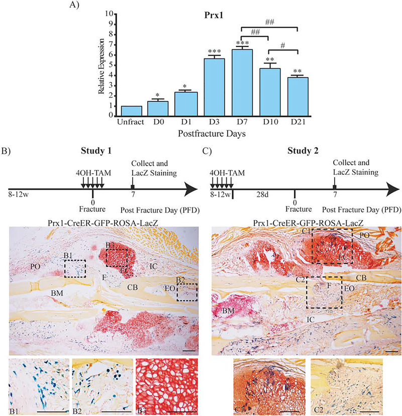
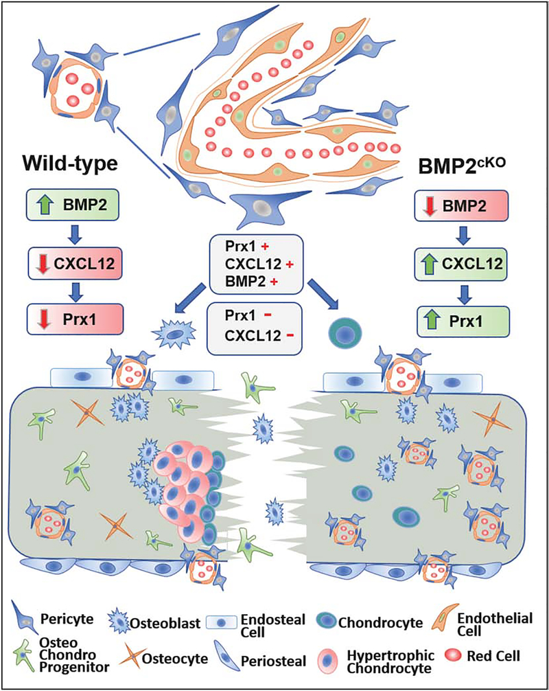
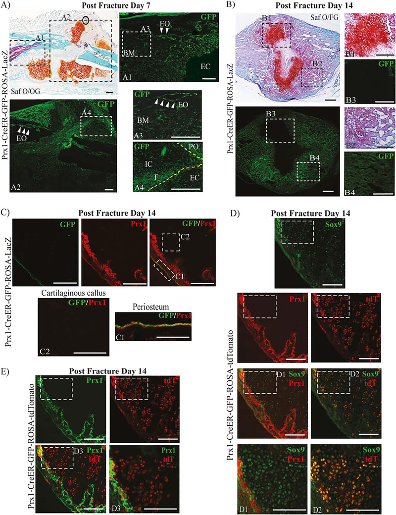
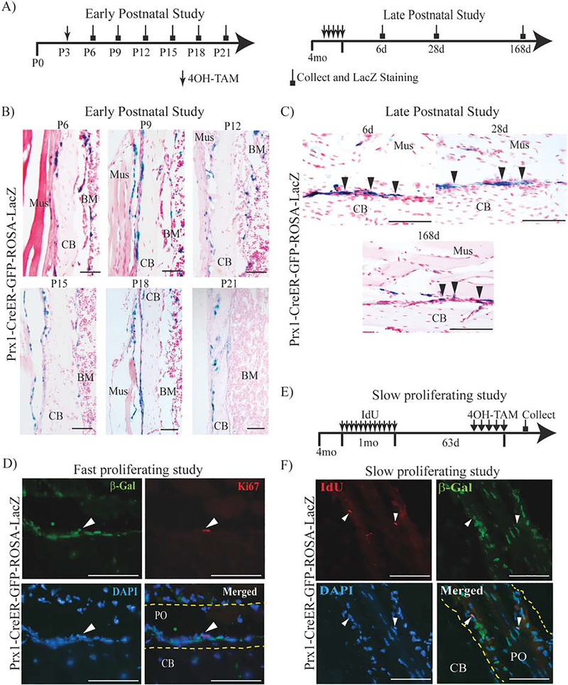
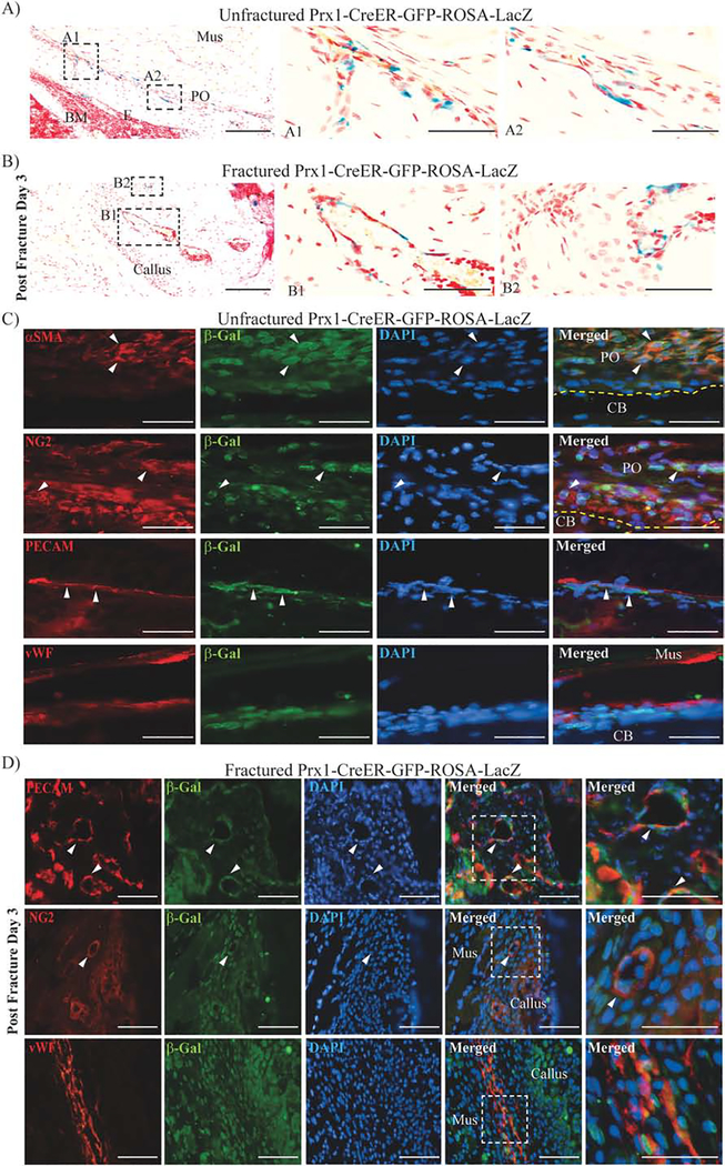
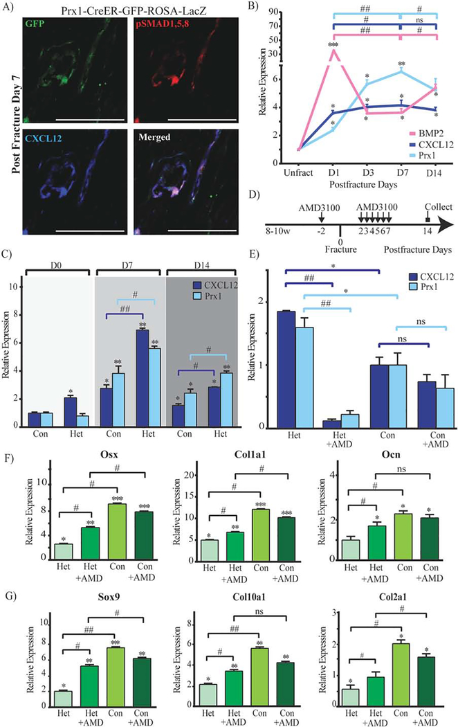
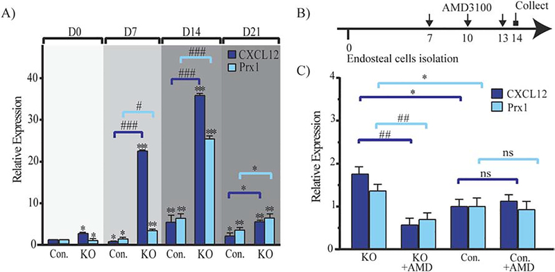
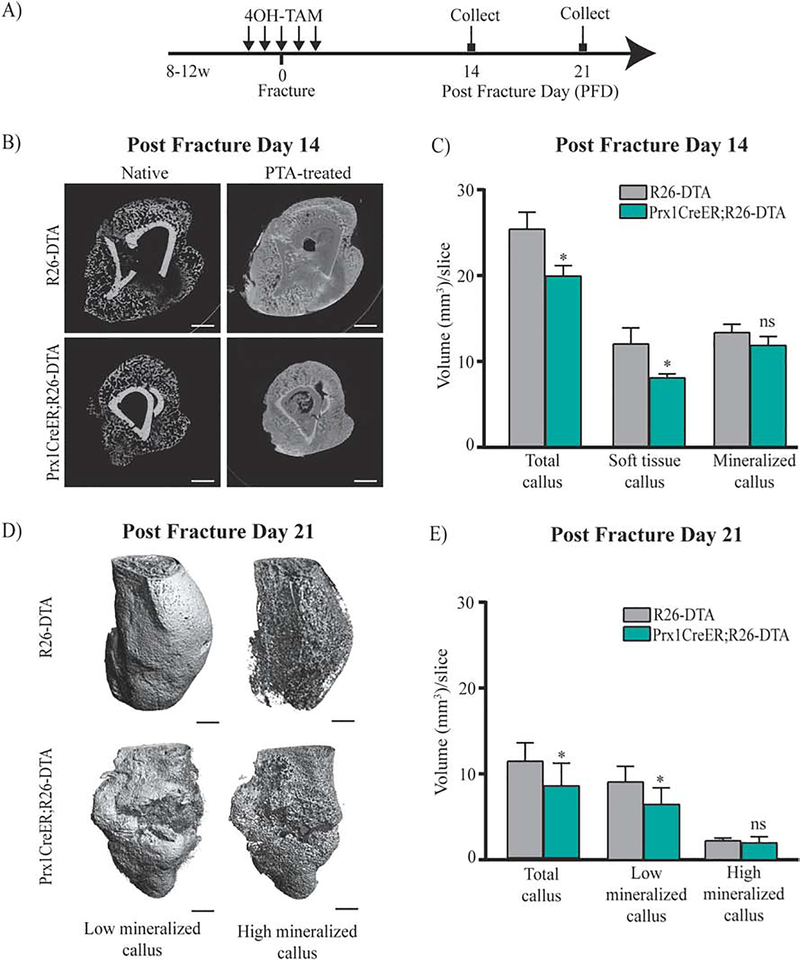
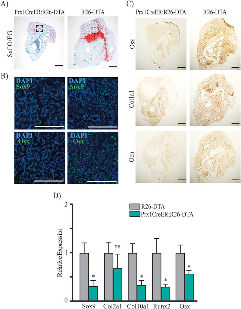
The healing capacity of bones after fracture implies the existence of adult regenerative cells. However, information on identification and functional role of fracture-induced progenitors is still lacking. Paired-related homeobox 1 (Prx1) is expressed during skeletogenesis. We hypothesize that fracture recapitulates Prx1's expression, and Prx1 expressing cells are critical to induce repair. To address our hypothesis, we used a combination of in vivo and in vitro approaches, short and long-term cell tracking analyses of progenies and actively expressing cells, cell ablation studies, and rodent animal models for normal and defective fracture healing. We found that fracture elicits a periosteal and endosteal response of perivascular Prx1+ cells that participate in fracture healing and showed that Prx1-expressing cells have a functional role in the repair process. While Prx1-derived cells contribute to the callus, Prx1's expression decreases concurrently with differentiation into cartilaginous and bone cells, similarly to when Prx1+ cells are cultured in differentiating conditions. We determined that bone morphogenic protein 2 (BMP2), through C-X-C motif-ligand-12 (CXCL12) signaling, modulates the downregulation of Prx1. We demonstrated that fracture elicits an early increase in BMP2 expression, followed by a decrease in CXCL12 that in turn down-regulates Prx1, allowing cells to commit to osteochondrogenesis. In vivo and in vitro treatment with CXCR4 antagonist AMD3100 restored Prx1 expression by modulating the BMP2-CXCL12 axis. Our studies represent a shift in the current research that has primarily focused on the identification of markers for postnatal skeletal progenitors, and instead we characterized the function of a specific population (Prx1+ cells) and their expression marker (Prx1) as a crossroad in fracture repair. The identification of fracture-induced perivascular Prx1+ cells and regulation of Prx1's expression by BMP2 and in turn by CXCL12 in the orchestration of fracture repair, highlights a pathway in which to investigate defective mechanisms and therapeutic targets for fracture non-union.

摘要

骨折后骨骼的愈合能力意味着成体再生细胞的存在。然而,关于骨折诱导祖细胞的鉴定及其功能作用的信息仍然缺乏。成对相关同源盒1(Prx1)在骨骼发生过程中表达。我们假设骨折会重现Prx1的表达,并且表达Prx1的细胞对诱导修复至关重要。为了验证我们的假设,我们结合了体内和体外方法、对后代和活跃表达细胞的短期和长期细胞追踪分析、细胞消融研究以及用于正常和缺陷性骨折愈合的啮齿动物模型。我们发现骨折会引发血管周围Prx1+细胞的骨膜和骨内膜反应,这些细胞参与骨折愈合,并表明表达Prx1的细胞在修复过程中具有功能作用。虽然源自Prx1的细胞有助于形成骨痂,但Prx1的表达会随着分化为软骨细胞和骨细胞而同时降低,这与在分化条件下培养Prx1+细胞时的情况类似。我们确定骨形态发生蛋白2(BMP2)通过C-X-C基序配体12(CXCL12)信号通路调节Prx1的下调。我们证明骨折会引发BMP2表达的早期增加,随后CXCL12减少,进而下调Prx1,使细胞能够进行骨软骨生成。用CXCR4拮抗剂AMD3100进行体内和体外治疗可通过调节BMP2-CXCL12轴恢复Prx1表达。我们的研究代表了当前研究的一个转变,目前的研究主要集中在鉴定出生后骨骼祖细胞的标志物,而我们则将特定群体(Prx1+细胞)及其表达标志物(Prx1)的功能表征为骨折修复的一个交叉点。骨折诱导的血管周围Prx1+细胞的鉴定以及BMP2进而通过CXCL12对Prx1表达的调节在骨折修复的协调中,突出了一条研究骨折不愈合缺陷机制和治疗靶点的途径。

https://cdn.ncbi.nlm.nih.gov/pmc/blobs/090e/7484205/93ae8cf256fa/nihms-1610269-f0009.jpg
https://cdn.ncbi.nlm.nih.gov/pmc/blobs/090e/7484205/4a9ae41c4b17/nihms-1610269-f0001.jpg
https://cdn.ncbi.nlm.nih.gov/pmc/blobs/090e/7484205/6b8c8386f544/nihms-1610269-f0002.jpg
https://cdn.ncbi.nlm.nih.gov/pmc/blobs/090e/7484205/1b1678a17b5a/nihms-1610269-f0003.jpg
https://cdn.ncbi.nlm.nih.gov/pmc/blobs/090e/7484205/4f4250c1b441/nihms-1610269-f0004.jpg
https://cdn.ncbi.nlm.nih.gov/pmc/blobs/090e/7484205/6c10bf158cf7/nihms-1610269-f0005.jpg
https://cdn.ncbi.nlm.nih.gov/pmc/blobs/090e/7484205/09de75f17ecc/nihms-1610269-f0006.jpg
https://cdn.ncbi.nlm.nih.gov/pmc/blobs/090e/7484205/c5e0209bc18d/nihms-1610269-f0007.jpg
https://cdn.ncbi.nlm.nih.gov/pmc/blobs/090e/7484205/5695996fb808/nihms-1610269-f0008.jpg
https://cdn.ncbi.nlm.nih.gov/pmc/blobs/090e/7484205/93ae8cf256fa/nihms-1610269-f0009.jpg

相似文献

1
Role of Prx1-expressing skeletal cells and Prx1-expression in fracture repair.表达Prx1的骨骼细胞及Prx1表达在骨折修复中的作用
Bone. 2020 Oct;139:115521. doi: 10.1016/j.bone.2020.115521. Epub 2020 Jul 3.
2
BMP2 Regulation of CXCL12 Cellular, Temporal, and Spatial Expression is Essential During Fracture Repair.BMP2 对 CXCL12 细胞、时间和空间表达的调节在骨折修复过程中是必不可少的。
J Bone Miner Res. 2015 Nov;30(11):2014-27. doi: 10.1002/jbmr.2548. Epub 2015 Jun 15.
3
Prx1-expressing cells contributing to fracture repair require primary cilia for complete healing in mice.表达 Prx1 的细胞有助于骨折修复,而这些细胞在小鼠中需要初级纤毛才能完全愈合。
Bone. 2021 Feb;143:115738. doi: 10.1016/j.bone.2020.115738. Epub 2020 Nov 11.
4
Periosteal cells are a major source of soft callus in bone fracture.骨膜细胞是骨折中软骨痂的主要来源。
J Bone Miner Metab. 2013 Jul;31(4):390-8. doi: 10.1007/s00774-013-0429-x. Epub 2013 Mar 12.
5
CXCR4 mediates the effects of IGF-1R signaling in rodent bone homeostasis and fracture repair.CXCR4 介导 IGF-1R 信号在啮齿动物骨稳态和骨折修复中的作用。
Bone. 2023 Jan;166:116600. doi: 10.1016/j.bone.2022.116600. Epub 2022 Nov 9.
6
Prx1 cell subpopulations identified in various tissues with diverse quiescence and activation ability following fracture and BMP2 stimulation.在骨折和BMP2刺激后,在各种组织中鉴定出具有不同静止和激活能力的Prx1细胞亚群。
Front Physiol. 2023 Jan 30;14:1106474. doi: 10.3389/fphys.2023.1106474. eCollection 2023.
7
IL-17RA Signaling in Prx1+ Mesenchymal Cells Influences Fracture Healing in Mice.Prx1+ 间充质细胞中的 IL-17RA 信号传导影响小鼠骨折愈合。
Int J Mol Sci. 2024 Mar 28;25(7):3751. doi: 10.3390/ijms25073751.
8
Periosteum progenitors could stimulate bone regeneration in aged murine bone defect model.骨膜祖细胞可刺激老年小鼠骨缺损模型中的骨再生。
J Cell Mol Med. 2020 Oct;24(20):12199-12210. doi: 10.1111/jcmm.15891. Epub 2020 Sep 15.
9
Post natal expression of Prx1 labels appendicular restricted progenitor cell populations of multiple tissues.Prx1 在产后表达,标记了多个组织的附肢限制祖细胞群体。
J Cell Physiol. 2022 May;237(5):2550-2560. doi: 10.1002/jcp.30728. Epub 2022 Mar 26.
10
Stromal cell-derived factor 1/CXCR4 signaling is critical for the recruitment of mesenchymal stem cells to the fracture site during skeletal repair in a mouse model.在小鼠模型的骨骼修复过程中,基质细胞衍生因子1/CXCR4信号通路对于间充质干细胞募集至骨折部位至关重要。
Arthritis Rheum. 2009 Mar;60(3):813-23. doi: 10.1002/art.24330.

引用本文的文献

1
Overactivation of EGFR signaling in skeletal stem/progenitor cells promotes bone formation and repair.骨骼干/祖细胞中表皮生长因子受体(EGFR)信号的过度激活可促进骨形成和修复。
Theranostics. 2025 Jul 24;15(16):8117-8136. doi: 10.7150/thno.115406. eCollection 2025.
2
Prx1 progenitors give rise to new articular cartilage when conditions are permissive for endogenous regeneration.当条件允许内源性再生时,Prx1祖细胞可产生新的关节软骨。
NPJ Regen Med. 2025 Aug 9;10(1):38. doi: 10.1038/s41536-025-00425-y.
3
+ fibroadipogenic progenitors in muscle are crucial for bone fracture repair.

本文引用的文献

1
American Society for Bone and Mineral Research-Orthopaedic Research Society Joint Task Force Report on Cell-Based Therapies.美国骨与矿物质研究学会 - 骨科学研究学会关于细胞疗法的联合特别工作组报告
J Bone Miner Res. 2020 Jan;35(1):3-17. doi: 10.1002/jbmr.3839. Epub 2019 Sep 23.
2
The limb enhancer marks an adult subpopulation of injury-responsive dermal fibroblasts.肢体增强子标记了成年期对损伤有反应的真皮成纤维细胞亚群。
Biol Open. 2019 Jul 18;8(7):bio043711. doi: 10.1242/bio.043711.
3
Prx1 Expressing Cells Are Required for Periodontal Regeneration of the Mouse Incisor.
肌肉中的纤维脂肪生成祖细胞对骨折修复至关重要。
Proc Natl Acad Sci U S A. 2025 Aug 5;122(31):e2417806122. doi: 10.1073/pnas.2417806122. Epub 2025 Jul 29.
4
Effects of aging on the immune and periosteal response to fracture injury.衰老对骨折损伤免疫及骨膜反应的影响。
Bone. 2025 Sep;198:117524. doi: 10.1016/j.bone.2025.117524. Epub 2025 May 15.
5
Prrx1-driven LINC complex disruption in vivo reduces osteoid deposition but not bone quality after voluntary wheel running.Prrx1 驱动的 LINC 复合物在体破坏减少了自发性跑步后的类骨质沉积,但不影响骨质量。
PLoS One. 2024 Nov 20;19(11):e0307816. doi: 10.1371/journal.pone.0307816. eCollection 2024.
6
Identification of Postn+ periosteal progenitor cells with bone regenerative potential.鉴定具有骨再生潜力的 Postn+骨膜前体细胞。
JCI Insight. 2024 Oct 8;9(19):e182524. doi: 10.1172/jci.insight.182524.
7
Neural regulation of mesenchymal stem cells in craniofacial bone: development, homeostasis and repair.间充质干细胞在颅面骨中的神经调节:发育、稳态与修复
Front Physiol. 2024 Jul 29;15:1423539. doi: 10.3389/fphys.2024.1423539. eCollection 2024.
8
Mechanosensitive protein polycystin-1 promotes periosteal stem/progenitor cells osteochondral differentiation in fracture healing.机械敏感蛋白多囊蛋白-1促进骨折愈合过程中骨膜干/祖细胞的骨软骨分化。
Theranostics. 2024 Apr 8;14(6):2544-2559. doi: 10.7150/thno.93269. eCollection 2024.
9
IL-17RA Signaling in Prx1+ Mesenchymal Cells Influences Fracture Healing in Mice.Prx1+ 间充质细胞中的 IL-17RA 信号传导影响小鼠骨折愈合。
Int J Mol Sci. 2024 Mar 28;25(7):3751. doi: 10.3390/ijms25073751.
10
Fracture healing research: Recent insights.骨折愈合研究:最新见解。
Bone Rep. 2023 May 19;19:101686. doi: 10.1016/j.bonr.2023.101686. eCollection 2023 Dec.
表达Prx1的细胞是小鼠切牙牙周再生所必需的。
Front Physiol. 2019 May 22;10:591. doi: 10.3389/fphys.2019.00591. eCollection 2019.
4
PDGF Modulates BMP2-Induced Osteogenesis in Periosteal Progenitor Cells.血小板衍生生长因子调节骨膜祖细胞中骨形态发生蛋白2诱导的成骨作用。
JBMR Plus. 2019 Jan 15;3(5):e10127. doi: 10.1002/jbm4.10127. eCollection 2019 May.
5
Mechanical Loading Promotes the Expansion of Primitive Osteoprogenitors and Organizes Matrix and Vascular Morphology in Long Bone Defects.机械负荷促进原始骨祖细胞的增殖并组织长骨缺损处的基质和血管形态。
J Bone Miner Res. 2019 May;34(5):896-910. doi: 10.1002/jbmr.3668. Epub 2019 Feb 20.
6
Relative contributions of adipose-resident CD146 pericytes and CD34 adventitial progenitor cells in bone tissue engineering.脂肪组织驻留的CD146周细胞和CD34外膜祖细胞在骨组织工程中的相对贡献
NPJ Regen Med. 2019 Jan 7;4:1. doi: 10.1038/s41536-018-0063-2. eCollection 2019.
7
Discovery of a periosteal stem cell mediating intramembranous bone formation.发现一种骨膜干细胞介导膜内成骨。
Nature. 2018 Oct;562(7725):133-139. doi: 10.1038/s41586-018-0554-8. Epub 2018 Sep 24.
8
Primary cilia are necessary for Prx1-expressing cells to contribute to postnatal skeletogenesis.初级纤毛对于 Prx1 表达细胞参与出生后骨骼发生是必需的。
J Cell Sci. 2018 Aug 20;131(16):jcs217828. doi: 10.1242/jcs.217828.
9
Periosteum contains skeletal stem cells with high bone regenerative potential controlled by Periostin.骨膜含有具有高骨再生潜力的骨骼干细胞,其受骨膜蛋白调控。
Nat Commun. 2018 Feb 22;9(1):773. doi: 10.1038/s41467-018-03124-z.
10
Stromal cell-derived factor-1α and transforming growth factor-β synergistically facilitate migration and chondrogenesis of synovium-derived stem cells through MAPK pathways.基质细胞衍生因子-1α与转化生长因子-β通过丝裂原活化蛋白激酶(MAPK)信号通路协同促进滑膜来源干细胞的迁移与软骨形成。
Am J Transl Res. 2017 May 15;9(5):2656-2667. eCollection 2017.