Department of Ophthalmology and Eye Hospital, University of Leipzig, Liebigstrasse 10-14, 04103, Leipzig, Germany.
Int Ophthalmol. 2020 Nov;40(11):2931-2948. doi: 10.1007/s10792-020-01477-3. Epub 2020 Jul 6.
To document with spectral-domain optical coherence tomography the structural stabilization of the fovea and the sealing of outer macular defects by Müller cells.
A retrospective case series of 45 eyes of 34 patients is described.
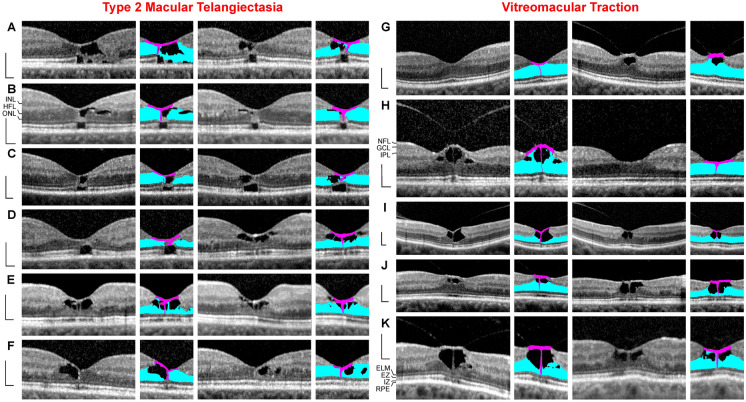
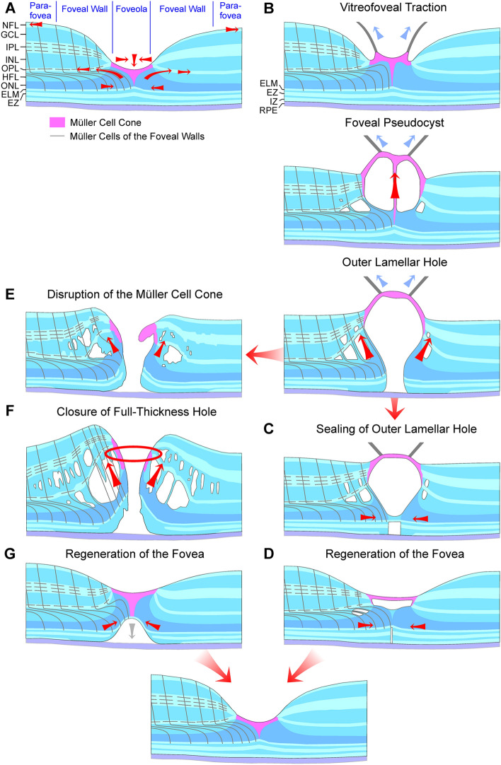
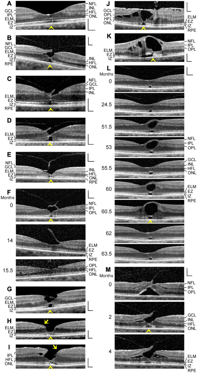
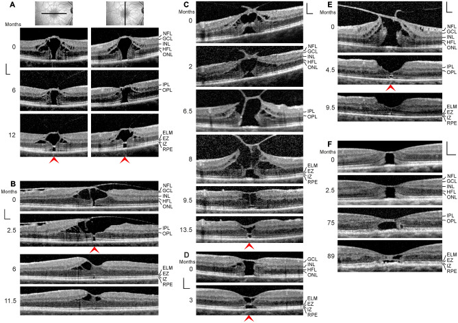
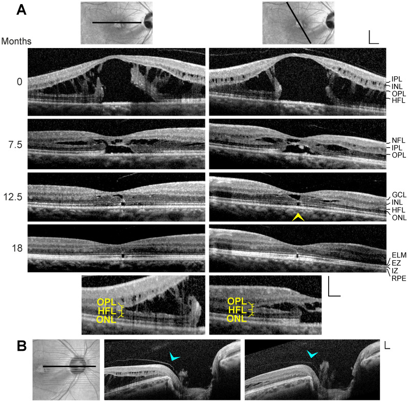
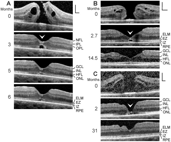
In cases of a cystic disruption of the foveola as in macular telangiectasia type 2 and vitreomacular traction, the Müller cell cone provides the structural stability of the fovea. In cases of a detachment or disruption of the Müller cell cone, e.g., in foveal pseudocysts, outer lamellar holes, and degenerative and tractional lamellar holes, Müller cells of the foveal walls may provide the structural stability of the fovea by the formation of a hyperreflective external limiting membrane (ELM) which bridges the holes in the central outer nuclear layer (ONL). Müller cells of the foveal walls and parafovea mediate the regeneration of the foveal architecture in cases of outer lamellar and full-thickness macular holes. The regeneration proceeds by a centripetal displacement of photoreceptor cell somata which closes the holes in the central ONL. The closure may be supported by the formation of a glial tissue band at the ELM which seals the hole.
The Müller cell cone provides the foveal stability in cases of a cystic disruption of the foveola. The structural stability of the outer foveal layers is mainly provided by the Müller cells of the foveal walls and parafovea; these cells also mediate the regeneration of the outer fovea in cases of a defect of the central ONL.
通过频域光相干断层扫描记录 Müller 细胞对黄斑中心凹的稳定作用及对外层黄斑裂孔的封闭作用。
回顾性病例系列研究,共纳入 34 例 45 只眼。
在 2 型黄斑毛细血管扩张症和玻璃体黄斑牵引中,黄斑中心凹小凹的囊样破坏由 Müller 细胞锥体提供结构稳定性。在外层板层孔、假性黄斑中心凹裂孔以及退行性和牵引性板层孔中,当 Müller 细胞锥体脱离或破坏时,黄斑中心凹壁的 Müller 细胞可能通过形成桥接中心外核层(ONL)中孔的高反射性外界膜(ELM)来提供中心凹的结构稳定性。在黄斑外层和全层裂孔中,Müller 细胞介导了黄斑中心凹结构的再生。该过程通过光感受器细胞体的向心性移位进行,从而封闭中心 ONL 中的孔。ELM 处胶质组织带的形成可能有助于封闭孔,支持其闭合。
在黄斑中心凹小凹的囊样破坏中,Müller 细胞锥体提供了中心凹的稳定性。外层黄斑中心凹的结构稳定性主要由黄斑中心凹壁和旁中心凹的 Müller 细胞提供;在中心 ONL 缺损的情况下,这些细胞也介导了外层黄斑的再生。