Division of Biomedical Engineering, School of Engineering, University of Glasgow, GlasgowG12 8LT, United Kingdom.
Bone and Joint Research Group, Centre for Human Development Stem Cells and Regeneration, University of Southampton, Southampton SO16 6YD, United Kingdom.
ACS Appl Mater Interfaces. 2020 Jul 29;12(30):33541-33549. doi: 10.1021/acsami.0c10273. Epub 2020 Jul 17.
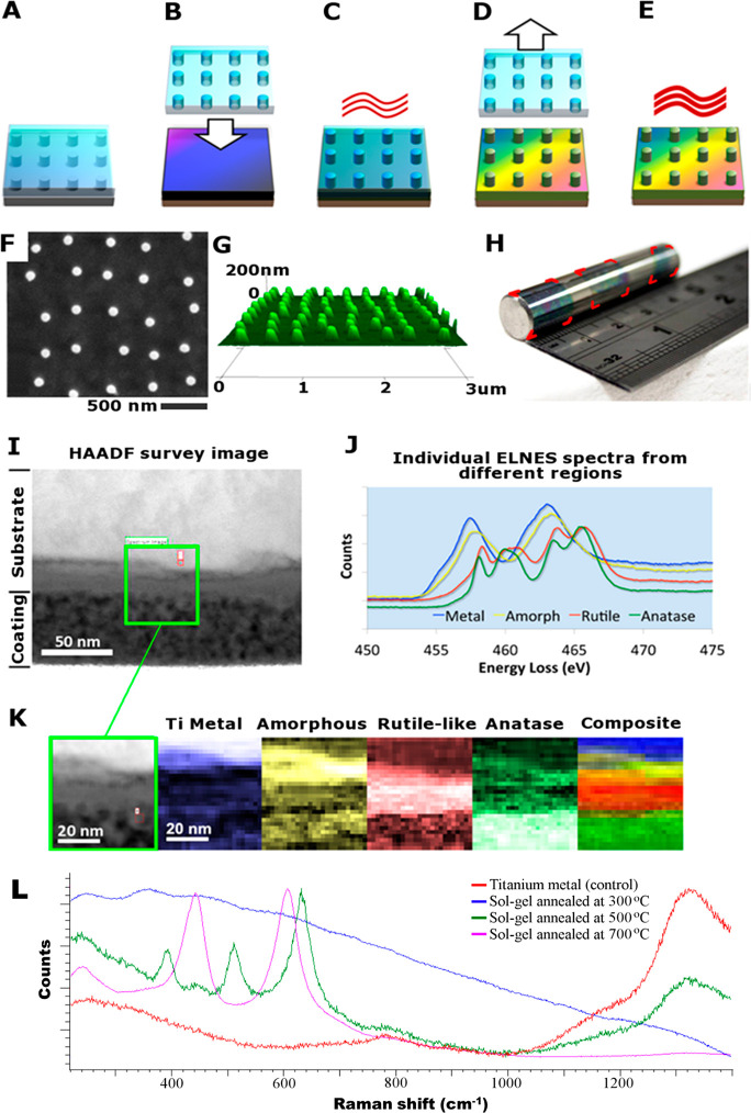
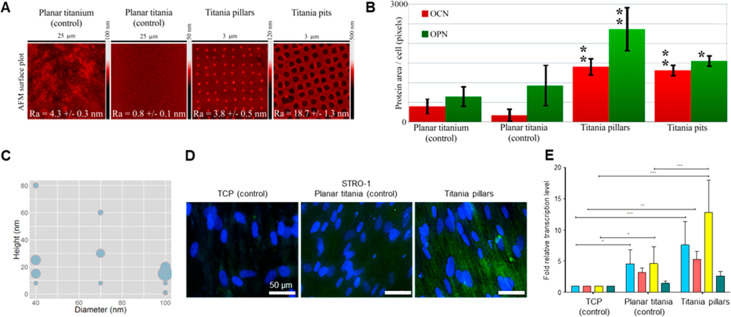
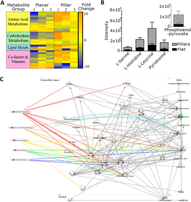
Accelerated de novo formation of bone is a highly desirable aim of implants targeting musculoskeletal injuries. To date, this has primarily been addressed by biologic factors. However, there is an unmet need for robust, highly reproducible yet economic alternative strategies that strongly induce an osteogenic cell response. Here, we present a surface engineering method of translating bioactive nanopatterns from polymeric in vitro studies to clinically relevant material for orthopedics: three-dimensional, large area metal. We use a titanium-based sol-gel whereby metal implants can be engineered to induce osteoinduction both in vitro and in vivo. We show that controlled disordered nanotopographies presented as pillars with 15-25 nm height and 100 nm diameter on titanium dioxide effectively induce osteogenesis when seeded with STRO-1-enriched human skeletal stem cells in vivo subcutaneous implantation in mice. After 28 days, samples were retrieved, which showed a 20-fold increase in osteogenic gene induction of nanopatterned substrates, indicating that the sol-gel nanopatterning method offers a promising route for translation to future clinical orthopedic implants.
加速新骨形成是治疗肌肉骨骼损伤的植入物的一个非常理想的目标。迄今为止,这主要是通过生物因素来实现的。然而,人们迫切需要一种强大、高度可重复但又经济的替代策略,这种策略能强烈诱导成骨细胞反应。在这里,我们提出了一种表面工程方法,即将生物活性纳米图案从聚合物的体外研究转化为临床相关的骨科材料:三维、大面积金属。我们使用基于钛的溶胶-凝胶,通过这种方法可以对金属植入物进行工程设计,以在体外和体内诱导成骨诱导。我们发现,当将富含 STRO-1 的人类骨骼干细胞接种在二氧化钛上的 15-25nm 高、100nm 直径的无序纳米柱形图案表面时,体内皮下植入小鼠后能有效地诱导成骨。28 天后,取出样本,结果显示纳米图案化基底的成骨基因诱导增加了 20 倍,这表明溶胶-凝胶纳米图案化方法为未来的临床骨科植入物提供了一种有前途的转化途径。