Cardiovascular Medicine Section, Department of Medicine, Boston University Medical Center, Boston, MA, USA.
Cardiovascular Research Center, Massachusetts General Hospital, Boston, MA, USA.
Sci Rep. 2020 Jul 8;10(1):11209. doi: 10.1038/s41598-020-68065-4.
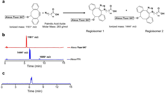
Multiplexed imaging is essential for the evaluation of substrate utilization in metabolically active organs, such as the heart and brown adipose tissue (BAT), where substrate preference changes in pathophysiologic states. Optical imaging provides a useful platform because of its low cost, high throughput and intrinsic ability to perform composite readouts. However, the paucity of probes available for in vivo use has limited optical methods to image substrate metabolism. Here, we present a novel near-infrared (NIR) free fatty acid (FFA) tracer suitable for in vivo imaging of deep tissues such as the heart. Using click chemistry, Alexa Fluor 647 DIBO Alkyne was conjugated to palmitic acid. Mice injected with 0.05 nmol/g bodyweight of the conjugate (AlexaFFA) were subjected to conditions known to increase FFA uptake in the heart (fasting) and BAT [cold exposure and injection with the β adrenergic agonist CL 316, 243(CL)]. Organs were subsequently imaged both ex vivo and in vivo to quantify AlexaFFA uptake. The blood kinetics of AlexaFFA followed a two-compartment model with an initial fast compartment half-life of 0.14 h and a subsequent slow compartment half-life of 5.2 h, consistent with reversible protein binding. Ex vivo fluorescence imaging after overnight cold exposure and fasting produced a significant increase in AlexaFFA uptake in the heart (58 ± 12%) and BAT (278 ± 19%) compared to warm/fed animals. In vivo imaging of the heart and BAT after exposure to CL and fasting showed a significant increase in AlexaFFA uptake in the heart (48 ± 20%) and BAT (40 ± 10%) compared to saline-injected/fed mice. We present a novel near-infrared FFA tracer, AlexaFFA, that is suitable for in vivo quantification of FFA metabolism and can be applied in the context of a low cost, high throughput, and multiplexed optical imaging platform.
多模式成像对于评估代谢活跃器官(如心脏和棕色脂肪组织(BAT))中的底物利用至关重要,因为在病理生理状态下,底物偏好会发生变化。光学成像是一种有用的平台,因为其成本低、通量高且具有内在的进行复合读出的能力。然而,由于体内可用的探针数量有限,光学方法仅限于对底物代谢进行成像。在这里,我们提出了一种新型的近红外(NIR)游离脂肪酸(FFA)示踪剂,适用于心脏等深部组织的体内成像。使用点击化学,将 Alexa Fluor 647 DIBO 炔烃与棕榈酸缀合。将 0.05 nmol/g 体重的缀合物(AlexaFFA)注射到小鼠体内,使其处于已知会增加心脏(禁食)和 BAT(冷暴露和注射β肾上腺素激动剂 CL 316,243(CL))中 FFA 摄取的条件下。随后,对器官进行离体和体内成像以定量摄取 AlexaFFA。AlexaFFA 的血液动力学遵循双室模型,初始快速隔室半衰期为 0.14 h,随后缓慢隔室半衰期为 5.2 h,与可逆蛋白结合一致。过夜冷暴露和禁食后的离体荧光成像导致 AlexaFFA 在心脏(58±12%)和 BAT(278±19%)中的摄取显著增加,与温暖/进食动物相比。暴露于 CL 和禁食后对心脏和 BAT 的体内成像显示,与盐水注射/进食的小鼠相比,AlexaFFA 在心脏(48±20%)和 BAT(40±10%)中的摄取显著增加。我们提出了一种新型的近红外 FFA 示踪剂 AlexaFFA,它适用于 FFA 代谢的体内定量,并可应用于低成本、高通量和多模式光学成像平台。