• 文献检索
  • 文档翻译
  • 深度研究
  • 学术资讯
  • Suppr Zotero 插件Zotero 插件
  • 邀请有礼
  • 套餐&价格
  • 历史记录
应用&插件
Suppr Zotero 插件Zotero 插件浏览器插件Mac 客户端Windows 客户端微信小程序
定价
高级版会员购买积分包购买API积分包
服务
文献检索文档翻译深度研究API 文档MCP 服务
关于我们
关于 Suppr公司介绍联系我们用户协议隐私条款
关注我们

Suppr 超能文献

核心技术专利:CN118964589B侵权必究
粤ICP备2023148730 号-1Suppr @ 2026

文献检索

告别复杂PubMed语法,用中文像聊天一样搜索,搜遍4000万医学文献。AI智能推荐,让科研检索更轻松。

立即免费搜索

文件翻译

保留排版,准确专业,支持PDF/Word/PPT等文件格式,支持 12+语言互译。

免费翻译文档

深度研究

AI帮你快速写综述,25分钟生成高质量综述,智能提取关键信息,辅助科研写作。

立即免费体验

脑内海绵状血管畸形是由 ADAMTS5 对 versican 的蛋白水解作用驱动的。

Cerebral cavernous malformations are driven by ADAMTS5 proteolysis of versican.

机构信息

Department of Medicine and Cardiovascular Institute, University of Pennsylvania, Philadelphia, PA.

Department of Molecular Genetics and Microbiology, Duke University School of Medicine, Durham, NC.

出版信息

J Exp Med. 2020 Oct 5;217(10). doi: 10.1084/jem.20200140.

DOI:10.1084/jem.20200140
PMID:32648916
原文链接:https://pmc.ncbi.nlm.nih.gov/articles/PMC7537394/
Abstract

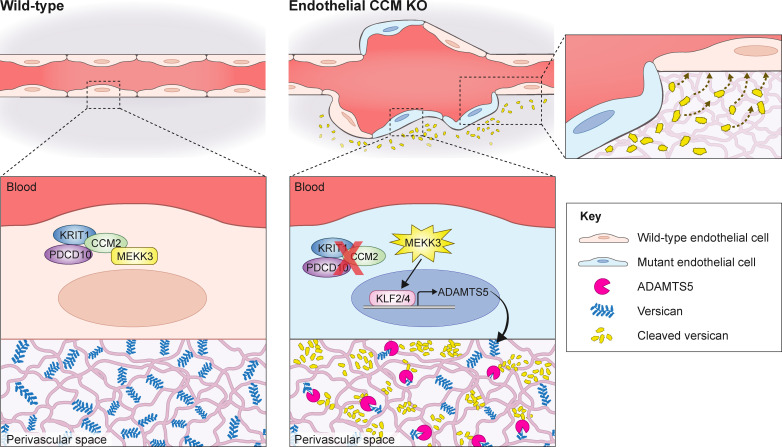
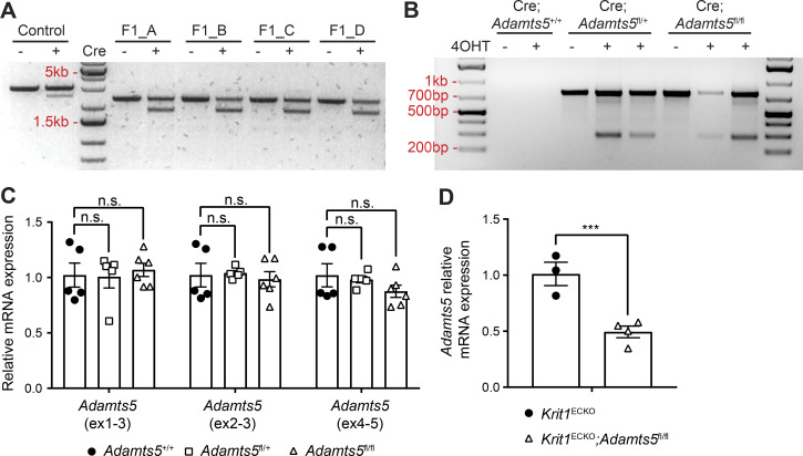
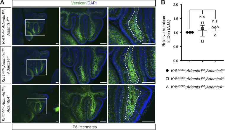
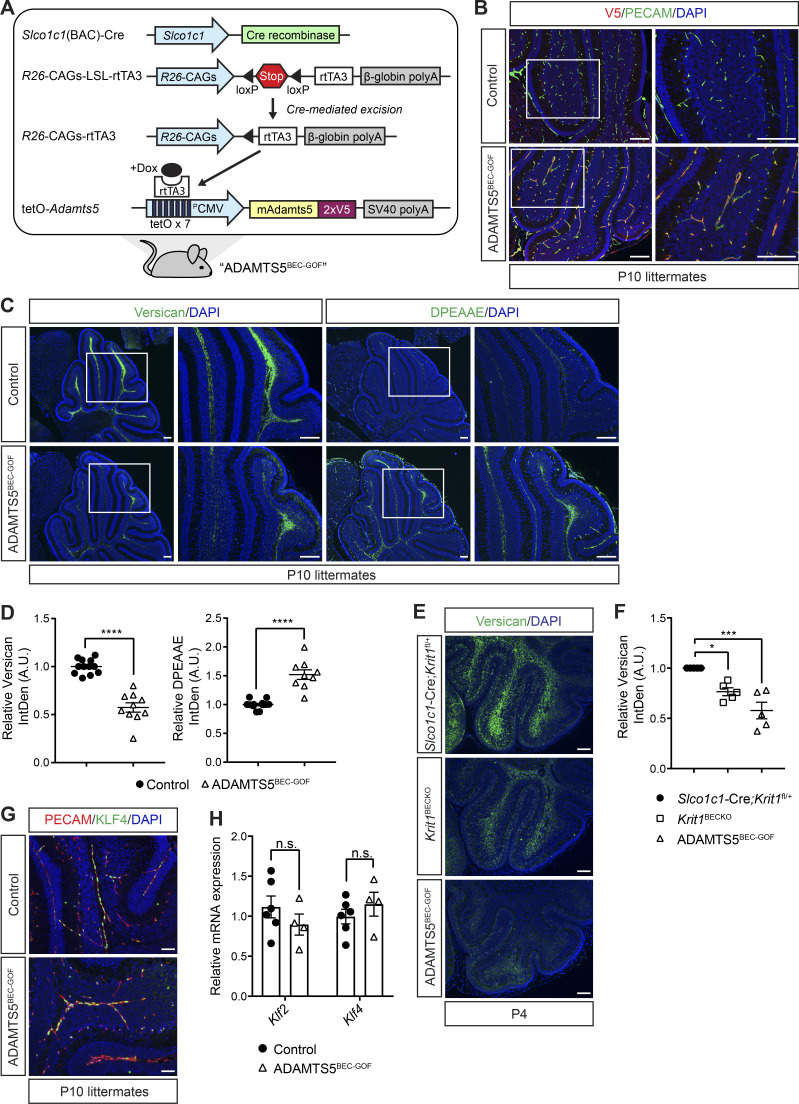
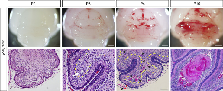
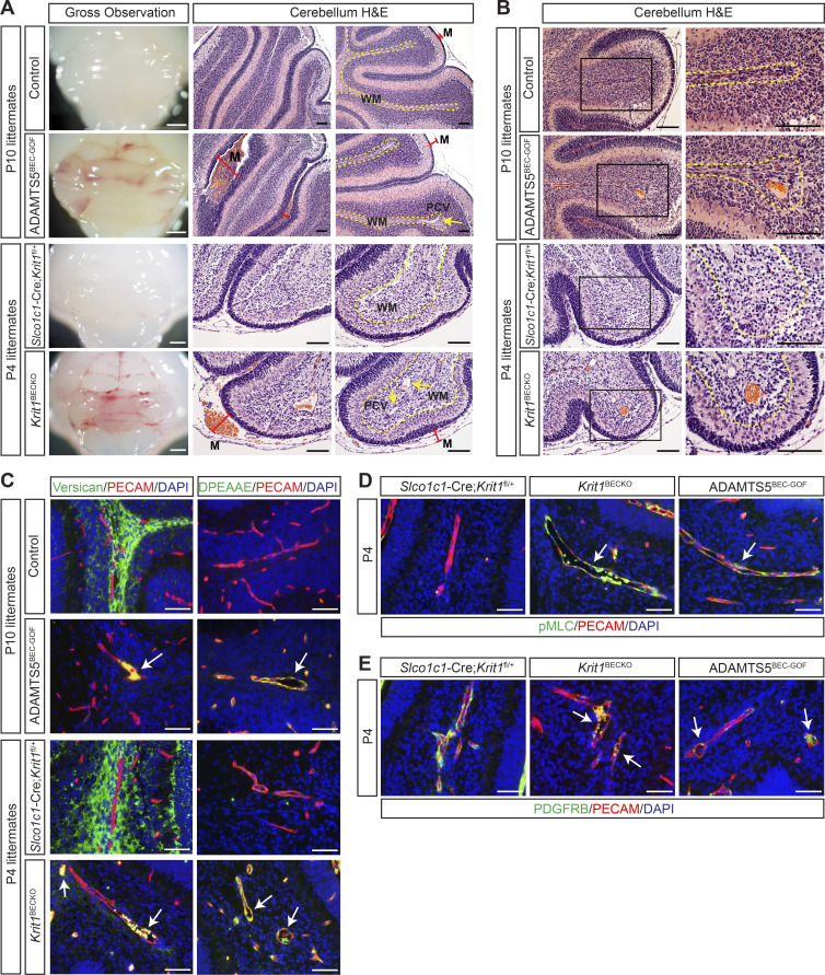
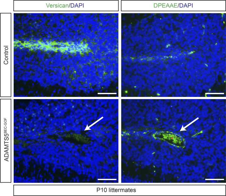
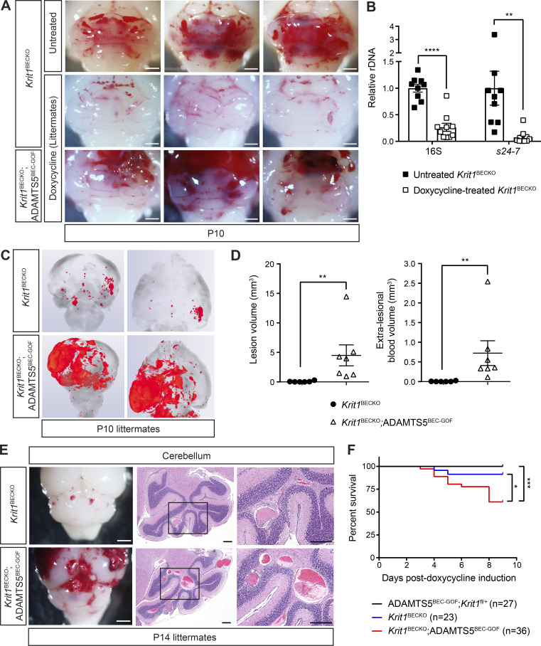
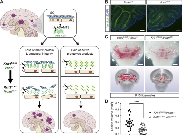
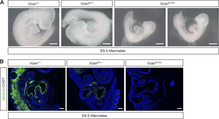
Cerebral cavernous malformations (CCMs) form following loss of the CCM protein complex in brain endothelial cells due to increased endothelial MEKK3 signaling and KLF2/4 transcription factor expression, but the downstream events that drive lesion formation remain undefined. Recent studies have revealed that CCM lesions expand by incorporating neighboring wild-type endothelial cells, indicative of a cell nonautonomous mechanism. Here we find that endothelial loss of ADAMTS5 reduced CCM formation in the neonatal mouse model. Conversely, endothelial gain of ADAMTS5 conferred early lesion genesis in the absence of increased KLF2/4 expression and synergized with KRIT1 loss of function to create large malformations. Lowering versican expression reduced CCM burden, indicating that versican is the relevant ADAMTS5 substrate and that lesion formation requires proteolysis but not loss of this extracellular matrix protein. These findings identify endothelial secretion of ADAMTS5 and cleavage of versican as downstream mechanisms of CCM pathogenesis and provide a basis for the participation of wild-type endothelial cells in lesion formation.

摘要

脑内海绵状血管畸形(CCMs)是由于脑内皮细胞中 CCM 蛋白复合物的丢失而形成的,这是由于内皮细胞 MEKK3 信号的增加和 KLF2/4 转录因子表达所致,但驱动病变形成的下游事件仍未定义。最近的研究表明,CCM 病变通过纳入相邻的野生型内皮细胞而扩张,表明存在细胞非自主性机制。在这里,我们发现内皮细胞中 ADAMTS5 的缺失减少了新生小鼠模型中的 CCM 形成。相反,内皮细胞中 ADAMTS5 的获得导致早期病变发生,而不增加 KLF2/4 的表达,并与 KRIT1 功能丧失协同作用,导致大的畸形。降低 versican 的表达减少了 CCM 的负担,表明 versican 是 ADAMTS5 的相关底物,病变的形成需要蛋白水解,但不需要这种细胞外基质蛋白的丢失。这些发现确定了内皮细胞分泌 ADAMTS5 和切割 versican 是 CCM 发病机制的下游机制,并为野生型内皮细胞参与病变形成提供了基础。

https://cdn.ncbi.nlm.nih.gov/pmc/blobs/c7cd/7537394/6446ea25129e/JEM_20200140_Fig7.jpg
https://cdn.ncbi.nlm.nih.gov/pmc/blobs/c7cd/7537394/896003ecbe40/JEM_20200140_GA.jpg
https://cdn.ncbi.nlm.nih.gov/pmc/blobs/c7cd/7537394/d2610f7c7397/JEM_20200140_Fig1.jpg
https://cdn.ncbi.nlm.nih.gov/pmc/blobs/c7cd/7537394/acd9197ba260/JEM_20200140_Fig2.jpg
https://cdn.ncbi.nlm.nih.gov/pmc/blobs/c7cd/7537394/ead78a7d542e/JEM_20200140_FigS1.jpg
https://cdn.ncbi.nlm.nih.gov/pmc/blobs/c7cd/7537394/2939b7f3636f/JEM_20200140_FigS2.jpg
https://cdn.ncbi.nlm.nih.gov/pmc/blobs/c7cd/7537394/f37388505b3a/JEM_20200140_Fig3.jpg
https://cdn.ncbi.nlm.nih.gov/pmc/blobs/c7cd/7537394/2ca56c3dc230/JEM_20200140_FigS3.jpg
https://cdn.ncbi.nlm.nih.gov/pmc/blobs/c7cd/7537394/f9eacf108be1/JEM_20200140_Fig4.jpg
https://cdn.ncbi.nlm.nih.gov/pmc/blobs/c7cd/7537394/4767fd92c01d/JEM_20200140_FigS4.jpg
https://cdn.ncbi.nlm.nih.gov/pmc/blobs/c7cd/7537394/3f63b02dd64b/JEM_20200140_Fig5.jpg
https://cdn.ncbi.nlm.nih.gov/pmc/blobs/c7cd/7537394/649b71b7e07d/JEM_20200140_Fig6.jpg
https://cdn.ncbi.nlm.nih.gov/pmc/blobs/c7cd/7537394/71442b63b77d/JEM_20200140_FigS5.jpg
https://cdn.ncbi.nlm.nih.gov/pmc/blobs/c7cd/7537394/6446ea25129e/JEM_20200140_Fig7.jpg

相似文献

1
Cerebral cavernous malformations are driven by ADAMTS5 proteolysis of versican.脑内海绵状血管畸形是由 ADAMTS5 对 versican 的蛋白水解作用驱动的。
J Exp Med. 2020 Oct 5;217(10). doi: 10.1084/jem.20200140.
2
Cerebral cavernous malformations arise from endothelial gain of MEKK3-KLF2/4 signalling.脑海绵状血管畸形源于MEKK3-KLF2/4信号通路的内皮细胞功能获得。
Nature. 2016 Apr 7;532(7597):122-6. doi: 10.1038/nature17178. Epub 2016 Mar 30.
3
Combined genetic-pharmacologic inactivation of tightly linked ADAMTS proteases in temporally specific windows uncovers distinct roles for versican proteolysis and glypican-6 in cardiac development.在特定时间窗口内联合遗传药理学失活紧密连锁的 ADAMTS 蛋白酶,揭示了 versican 蛋白水解和 glypican-6 在心脏发育中的不同作用。
Matrix Biol. 2024 Aug;131:1-16. doi: 10.1016/j.matbio.2024.05.003. Epub 2024 May 13.
4
ADAMTS4 and ADAMTS5 knockout mice are protected from versican but not aggrecan or brevican proteolysis during spinal cord injury.在脊髓损伤期间,ADAMTS4和ADAMTS5基因敲除小鼠可免受多功能蛋白聚糖的影响,但不能免受聚集蛋白聚糖或短蛋白聚糖的蛋白水解作用。
Biomed Res Int. 2014;2014:693746. doi: 10.1155/2014/693746. Epub 2014 Jul 3.
5
The C-terminal domains of ADAMTS1 contain exosites involved in its proteoglycanase activity.ADAMTS1 的 C 端结构域包含参与蛋白聚糖酶活性的外显子。
J Biol Chem. 2023 Apr;299(4):103048. doi: 10.1016/j.jbc.2023.103048. Epub 2023 Feb 21.
6
Role of ADAMTS-5 in Aortic Dilatation and Extracellular Matrix Remodeling.ADAMTS-5 在主动脉扩张和细胞外基质重塑中的作用。
Arterioscler Thromb Vasc Biol. 2018 Jul;38(7):1537-1548. doi: 10.1161/ATVBAHA.117.310562. Epub 2018 Apr 5.
7
Determinants of versican-V1 proteoglycan processing by the metalloproteinase ADAMTS5.金属蛋白酶ADAMTS5对多功能蛋白聚糖-V1蛋白聚糖进行加工处理的决定因素。
J Biol Chem. 2014 Oct 3;289(40):27859-73. doi: 10.1074/jbc.M114.573287. Epub 2014 Aug 13.
8
CDC42 Deletion Elicits Cerebral Vascular Malformations via Increased MEKK3-Dependent KLF4 Expression.CDC42 缺失通过增加 MEKK3 依赖性 KLF4 表达引发脑血管畸形。
Circ Res. 2019 Apr 12;124(8):1240-1252. doi: 10.1161/CIRCRESAHA.118.314300.
9
Cerebral cavernous malformations form an anticoagulant vascular domain in humans and mice.脑内海绵状血管畸形在人类和小鼠中形成一个抗凝血的血管区域。
Blood. 2019 Jan 17;133(3):193-204. doi: 10.1182/blood-2018-06-856062. Epub 2018 Nov 15.
10
Generation of CCM Phenotype by a Human Microvascular Endothelial Model.通过人微血管内皮细胞模型生成CCM表型
Methods Mol Biol. 2020;2152:131-137. doi: 10.1007/978-1-0716-0640-7_10.

引用本文的文献

1
Common and distinct circulating microRNAs in four neurovascular disorders.四种神经血管疾病中常见和独特的循环微小RNA
Biochem Biophys Rep. 2025 Aug 2;43:102189. doi: 10.1016/j.bbrep.2025.102189. eCollection 2025 Sep.
2
Focused ultrasound augments the delivery and penetration of model therapeutics into cerebral cavernous malformations.聚焦超声增强了模型治疗药物向脑海绵状血管畸形的递送和渗透。
J Control Release. 2025 Jul 10;383:113861. doi: 10.1016/j.jconrel.2025.113861. Epub 2025 May 16.
3
A Systematic Review of MicroRNAs in Hemorrhagic Neurovascular Disease: Cerebral Cavernous Malformations as a Paradigm.

本文引用的文献

1
Distinct cellular roles for PDCD10 define a gut-brain axis in cerebral cavernous malformation.PDCD10 在不同细胞中的作用定义了脑动静脉畸形的肠脑轴。
Sci Transl Med. 2019 Nov 27;11(520). doi: 10.1126/scitranslmed.aaw3521.
2
Mice Exhibit Altered Aggrecan Proteolytic Profiles That Correlate With Ascending Aortic Anomalies.老鼠表现出与升主动脉异常相关的改变的聚集蛋白聚糖蛋白水解谱。
Arterioscler Thromb Vasc Biol. 2019 Oct;39(10):2067-2081. doi: 10.1161/ATVBAHA.119.313077. Epub 2019 Aug 1.
3
Exosites in Hypervariable Loops of ADAMTS Spacer Domains control Substrate Recognition and Proteolysis.
出血性神经血管疾病中微小RNA的系统评价:以脑海绵状血管畸形为例
Int J Mol Sci. 2025 Apr 17;26(8):3794. doi: 10.3390/ijms26083794.
4
Focused Ultrasound Augments the Delivery and Penetration of Model Therapeutics into Cerebral Cavernous Malformations.聚焦超声增强模型治疗药物向脑海绵状血管畸形的递送与穿透。
bioRxiv. 2025 Jan 23:2024.08.27.609060. doi: 10.1101/2024.08.27.609060.
5
Hyaluronic acid turnover controls the severity of cerebral cavernous malformations in bioengineered human micro-vessels.透明质酸周转控制生物工程化人类微血管中脑海绵状血管畸形的严重程度。
APL Bioeng. 2024 Feb 12;8(1):016108. doi: 10.1063/5.0159330. eCollection 2024 Mar.
6
Identification of the Shared Gene Signatures of HCK, NOG, RNF125 and Biological Mechanism in Pediatric Acute Lymphoblastic Leukaemia and Pediatric Sepsis.小儿急性淋巴细胞白血病和小儿脓毒症中HCK、NOG、RNF125共享基因特征的鉴定及生物学机制
Mol Biotechnol. 2025 Jan;67(1):80-90. doi: 10.1007/s12033-023-00979-6. Epub 2023 Dec 20.
7
mTORC1 Inhibitor Rapamycin Inhibits Growth of Cerebral Cavernous Malformation in Adult Mice.mTORC1 抑制剂雷帕霉素抑制成年小鼠脑内海绵状血管畸形的生长。
Stroke. 2023 Nov;54(11):2906-2917. doi: 10.1161/STROKEAHA.123.044108. Epub 2023 Sep 25.
8
Plasma metabolites with mechanistic and clinical links to the neurovascular disease cavernous angioma.与神经血管疾病海绵状血管瘤存在机制和临床关联的血浆代谢物。
Commun Med (Lond). 2023 Mar 3;3(1):35. doi: 10.1038/s43856-023-00265-1.
9
Cell-autonomous requirement for ACE2 across organs in lethal mouse SARS-CoV-2 infection.器官中 ACE2 对致死性 SARS-CoV-2 感染的细胞自主需求。
PLoS Biol. 2023 Feb 6;21(2):e3001989. doi: 10.1371/journal.pbio.3001989. eCollection 2023 Feb.
10
Release of STK24/25 suppression on MEKK3 signaling in endothelial cells confers cerebral cavernous malformation.STK24/25 对内皮细胞中 MEKK3 信号的释放赋予了脑海绵状血管畸形。
JCI Insight. 2023 Mar 8;8(5):e160372. doi: 10.1172/jci.insight.160372.
ADAMTS 间隔域超变环中的外显子控制底物识别和蛋白水解。
Sci Rep. 2019 Jul 29;9(1):10914. doi: 10.1038/s41598-019-47494-w.
4
Endothelial cell clonal expansion in the development of cerebral cavernous malformations.脑动静脉畸形发生中内皮细胞的克隆性扩张。
Nat Commun. 2019 Jun 24;10(1):2761. doi: 10.1038/s41467-019-10707-x.
5
Comprehensive transcriptome analysis of cerebral cavernous malformation across multiple species and genotypes.跨多种物种和基因型的脑海绵状血管畸形的综合转录组分析。
JCI Insight. 2019 Feb 7;4(3):e126167. doi: 10.1172/jci.insight.126167.
6
Cerebral Cavernous Malformations Develop Through Clonal Expansion of Mutant Endothelial Cells.脑内海绵状血管畸形通过突变内皮细胞的克隆扩张发展。
Circ Res. 2018 Oct 26;123(10):1143-1151. doi: 10.1161/CIRCRESAHA.118.313970.
7
Control of cardiac jelly dynamics by NOTCH1 and NRG1 defines the building plan for trabeculation.NOTCH1 和 NRG1 通过控制心胶动力学来定义小梁化的构建计划。
Nature. 2018 May;557(7705):439-445. doi: 10.1038/s41586-018-0110-6. Epub 2018 May 9.
8
Role of ADAMTS-5 in Aortic Dilatation and Extracellular Matrix Remodeling.ADAMTS-5 在主动脉扩张和细胞外基质重塑中的作用。
Arterioscler Thromb Vasc Biol. 2018 Jul;38(7):1537-1548. doi: 10.1161/ATVBAHA.117.310562. Epub 2018 Apr 5.
9
Cerebral Cavernous Malformations: An Update on Prevalence, Molecular Genetic Analyses, and Genetic Counselling.脑海绵状血管畸形:患病率、分子遗传学分析及遗传咨询的最新进展
Mol Syndromol. 2018 Feb;9(2):60-69. doi: 10.1159/000486292. Epub 2018 Jan 25.
10
Massive aggrecan and versican accumulation in thoracic aortic aneurysm and dissection.胸主动脉瘤和夹层中大量聚集的聚集蛋白聚糖和软骨寡聚基质蛋白。
JCI Insight. 2018 Mar 8;3(5):97167. doi: 10.1172/jci.insight.97167.