Yordanov Teodor E, Keyser Mikaela S, Enriquez Martinez Marco A, Esposito Tyron, Tefft Juliann B, Morris Elysse K, Labzin Larisa I, Stehbens Samantha J, Rowan Alan E, Hogan Benjamin M, Chen Christopher S, Lauko Jan, Lagendijk Anne K
Centre for Cell Biology and Chronic Disease, Institute for Molecular Bioscience, The University of Queensland, Brisbane, Queensland, Australia.
Australian Institute for Bioengineering and Nanotechnology, The University of Queensland, Brisbane, Queensland, Australia.
APL Bioeng. 2024 Feb 12;8(1):016108. doi: 10.1063/5.0159330. eCollection 2024 Mar.
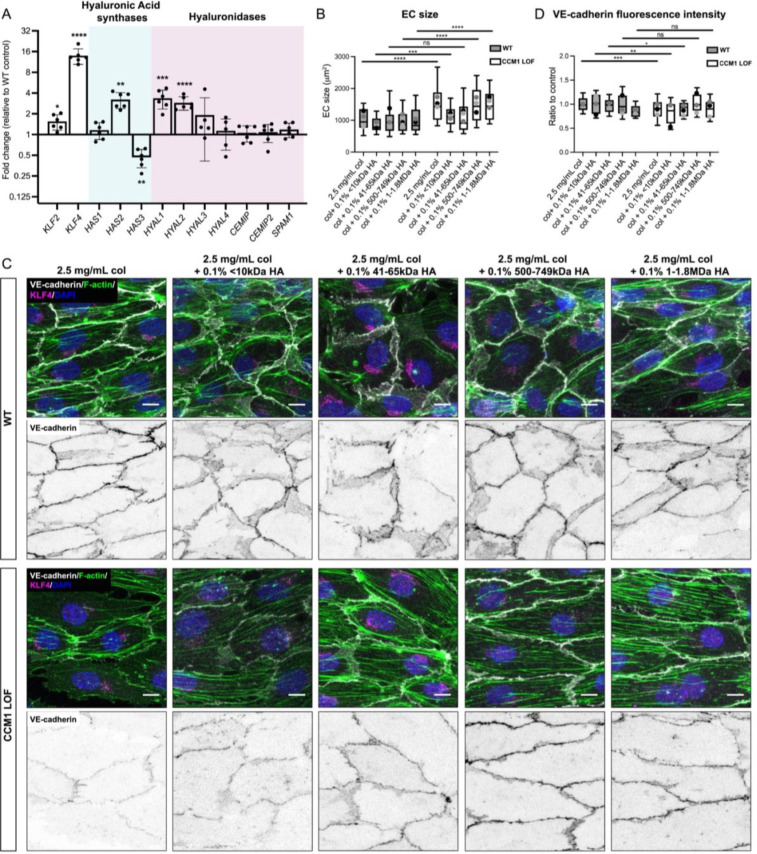
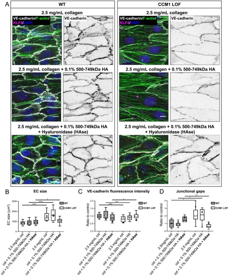
Cerebral cavernous malformations (CCMs) are vascular lesions that predominantly form in blood vessels of the central nervous system upon loss of the CCM multimeric protein complex. The endothelial cells within CCM lesions are characterized by overactive MEKK3 kinase and KLF2/4 transcription factor signaling, leading to pathological changes such as increased endothelial cell spreading and reduced junctional integrity. Concomitant to aberrant endothelial cell signaling, non-autonomous signals from the extracellular matrix (ECM) have also been implicated in CCM lesion growth and these factors might explain why CCM lesions mainly develop in the central nervous system. Here, we adapted a three-dimensional microfluidic system to examine CCM1 deficient human micro-vessels in distinctive extracellular matrices. We validate that pathological hallmarks are maintained in this model. We further show that key genes responsible for homeostasis of hyaluronic acid, a major extracellular matrix component of the central nervous system, are dysregulated in CCM. Supplementing the matrix in our model with distinct forms of hyaluronic acid inhibits pathological cell spreading and rescues barrier function. Hyaluronic acid acts by dampening cell-matrix adhesion signaling in CCM, either downstream or in parallel of KLF2/4. This study provides a proof-of-principle that ECM embedded 3D microfluidic models are ideally suited to identify how changes in ECM structure and signaling impact vascular malformations.
脑海绵状血管畸形(CCM)是一种血管病变,主要在CCM多聚体蛋白复合物缺失时于中枢神经系统的血管中形成。CCM病变内的内皮细胞具有过度活跃的MEKK3激酶和KLF2/4转录因子信号特征,导致诸如内皮细胞铺展增加和连接完整性降低等病理变化。与异常的内皮细胞信号传导同时,细胞外基质(ECM)的非自主信号也与CCM病变生长有关,这些因素可能解释了为什么CCM病变主要发生在中枢神经系统。在这里,我们采用了一种三维微流控系统来检测在不同细胞外基质中的CCM1缺陷型人类微血管。我们验证了该模型中病理特征得以维持。我们进一步表明,负责透明质酸(中枢神经系统主要细胞外基质成分)稳态的关键基因在CCM中失调。用不同形式的透明质酸补充我们模型中的基质可抑制病理细胞铺展并恢复屏障功能。透明质酸通过减弱CCM中细胞 - 基质黏附信号发挥作用,该作用发生在KLF2/4的下游或与其平行。本研究提供了一个原理证明,即嵌入细胞外基质的三维微流控模型非常适合确定细胞外基质结构和信号变化如何影响血管畸形。