Department of Psychosis Studies, Institute of Psychiatry, Psychology and Neuroscience, King's College London, London, United Kingdom; Psychiatric Imaging Group, MRC London Institute of Medical Sciences, Hammersmith Hospital, Imperial College London, London, United Kingdom; Institute of Clinical Sciences, Faculty of Medicine, Imperial College London, London, United Kingdom.
Department of Psychosis Studies, Institute of Psychiatry, Psychology and Neuroscience, King's College London, London, United Kingdom; Psychiatric Imaging Group, MRC London Institute of Medical Sciences, Hammersmith Hospital, Imperial College London, London, United Kingdom; Institute of Clinical Sciences, Faculty of Medicine, Imperial College London, London, United Kingdom.
Biol Psychiatry Cogn Neurosci Neuroimaging. 2020 Nov;5(11):1040-1051. doi: 10.1016/j.bpsc.2020.04.004. Epub 2020 Apr 23.
Striatal dopamine dysfunction is thought to underlie symptoms in psychosis, yet it remains unclear how a single neurotransmitter could cause the diverse presentations that are observed clinically. One hypothesis is that the consequences of aberrant dopamine signaling vary depending on where within the striatum the dysfunction occurs. Positron emission tomography allows for the quantification of dopamine function across the striatum. In the current study, we used a novel method to investigate the relationship between spatial variability in dopamine synthesis capacity and psychotic symptoms.
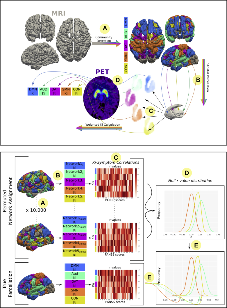
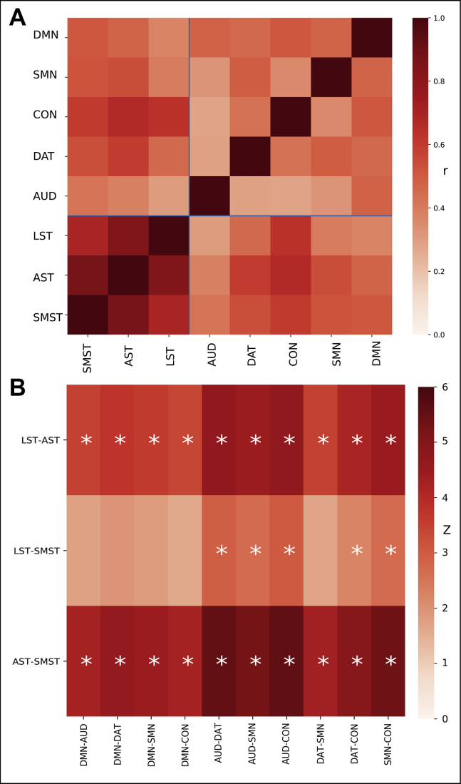
We used a multimodal imaging approach combining F-DOPA positron emission tomography and resting-state magnetic resonance imaging in 29 patients with first-episode psychosis and 21 healthy control subjects. In each participant, resting-state functional connectivity maps were used to quantify the functional connectivity of each striatal voxel to well-established cortical networks. Network-specific striatal dopamine synthesis capacity (Ki) was then calculated for the resulting connectivity-defined parcellations.
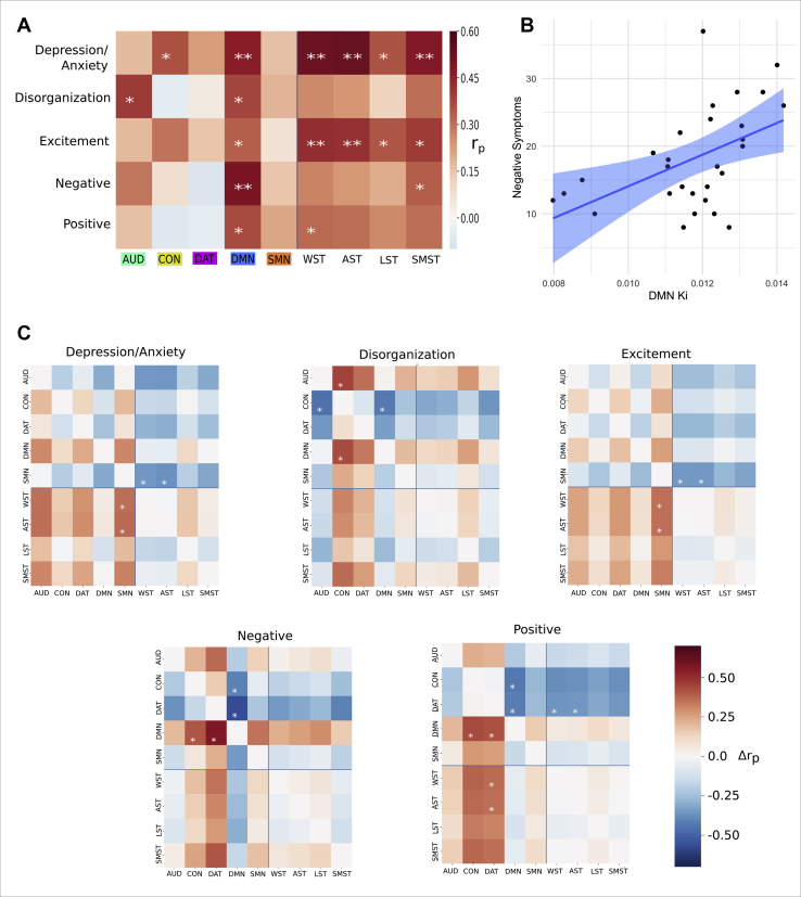
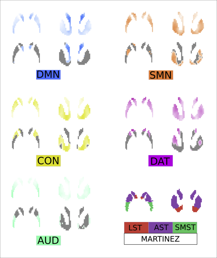
The connectivity-defined parcellations generated Ki values with equivalent reliability, and significantly greater orthogonality compared with standard anatomical parcellation methods. As a result, dopamine-symptom associations were significantly different from one another for different subdivisions, whereas no unique subdivision relationships were found when using an anatomical parcellation. In particular, dopamine function within striatal areas connected to the default mode network was strongly associated with negative symptoms (p < .001).
These findings suggest that individual differences in the topography of dopamine dysfunction within the striatum contribute to shaping psychotic symptomatology. Further validation of the novel approach in future studies is necessary.
纹状体多巴胺功能障碍被认为是精神分裂症症状的基础,但目前尚不清楚单一神经递质如何导致临床上观察到的各种表现。一种假设是,异常多巴胺信号传递的后果取决于纹状体中功能障碍发生的位置。正电子发射断层扫描允许在纹状体中量化多巴胺功能。在目前的研究中,我们使用一种新方法来研究多巴胺合成能力的空间变异性与精神病症状之间的关系。
我们使用一种结合 F-DOPA 正电子发射断层扫描和静息态磁共振成像的多模态成像方法,对 29 名首发精神分裂症患者和 21 名健康对照进行研究。在每个参与者中,使用静息态功能连接图来量化每个纹状体体素与既定皮质网络的功能连接。然后,为所得连接定义的分割计算特定于网络的纹状体多巴胺合成能力 (Ki)。
连接定义的分割产生了具有等效可靠性的 Ki 值,并且与标准解剖分割方法相比,具有显著更高的正交性。因此,多巴胺-症状之间的关联在不同的细分中明显不同,而使用解剖分割时则没有发现独特的细分关系。特别是,与默认模式网络相连的纹状体区域内的多巴胺功能与阴性症状强烈相关 (p<.001)。
这些发现表明,纹状体中多巴胺功能障碍的拓扑个体差异有助于塑造精神病症状。需要在未来的研究中进一步验证新方法。