Weyers Brent W, Marsden Mark, Sun Tianchen, Bec Julien, Bewley Arnaud F, Gandour-Edwards Regina F, Moore Michael G, Farwell D Gregory, Marcu Laura
Department of Biomedical Engineering, University of California, Davis, California.
Department of Computer Science, University of California, Davis, California.
Transl Biophotonics. 2019 Dec;1(1-2). doi: 10.1002/tbio.201900017. Epub 2019 Oct 29.
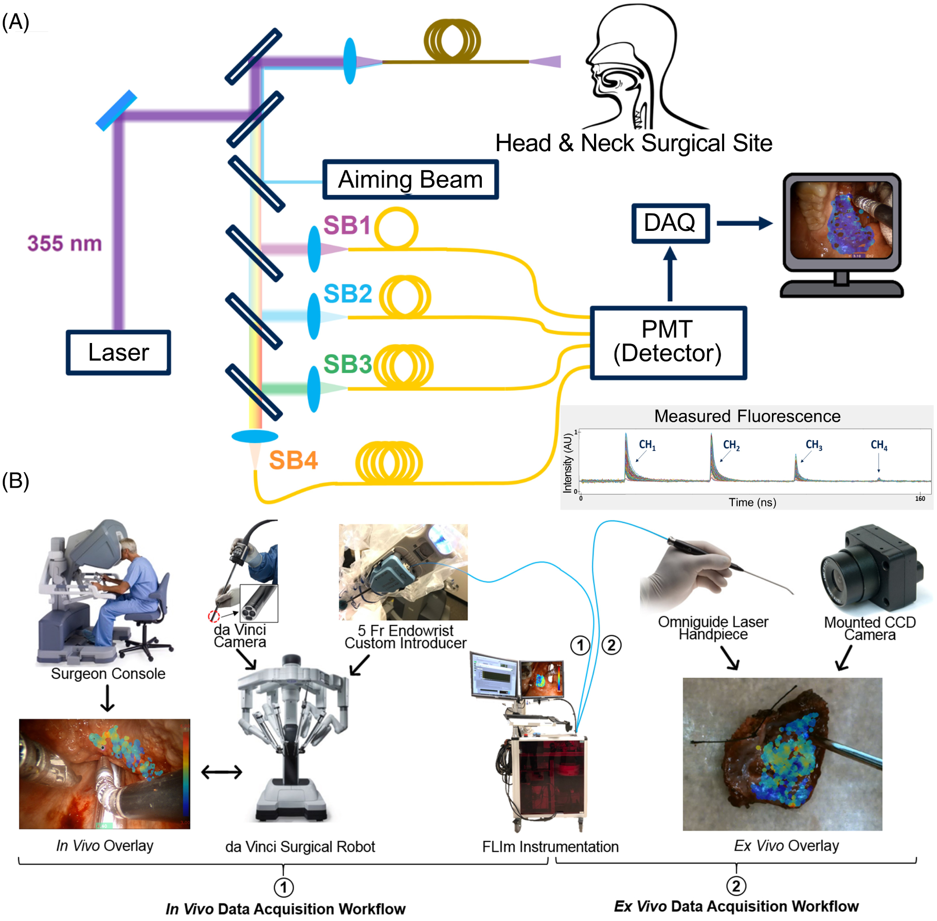
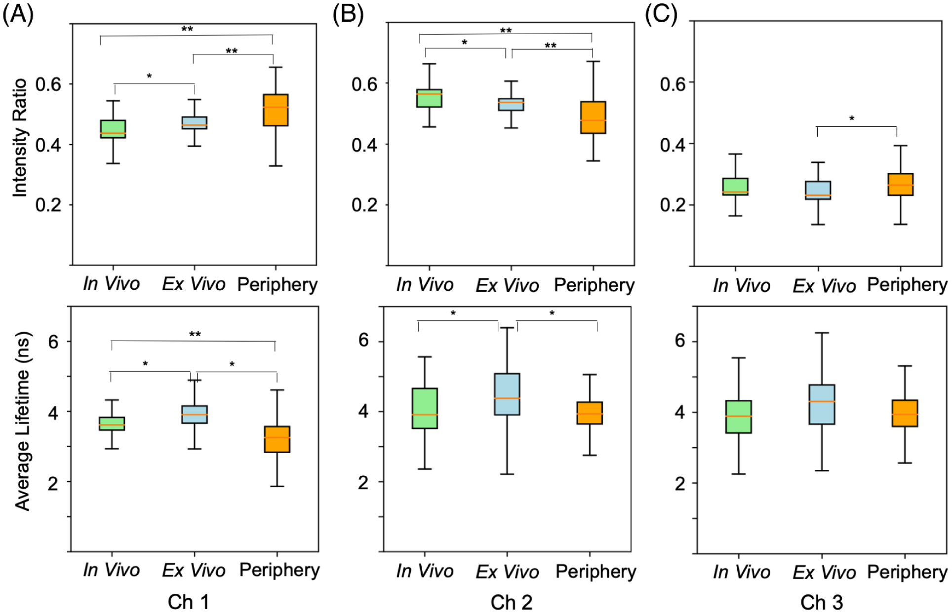
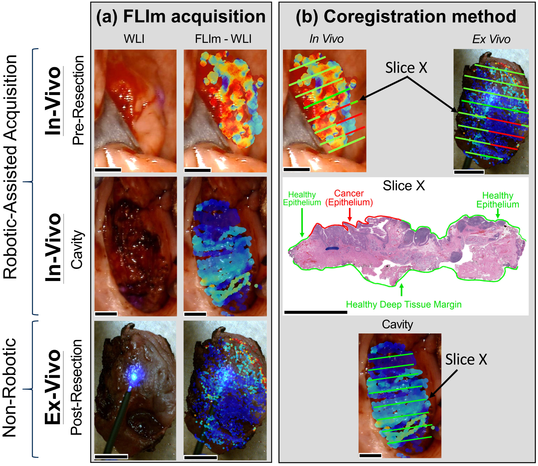
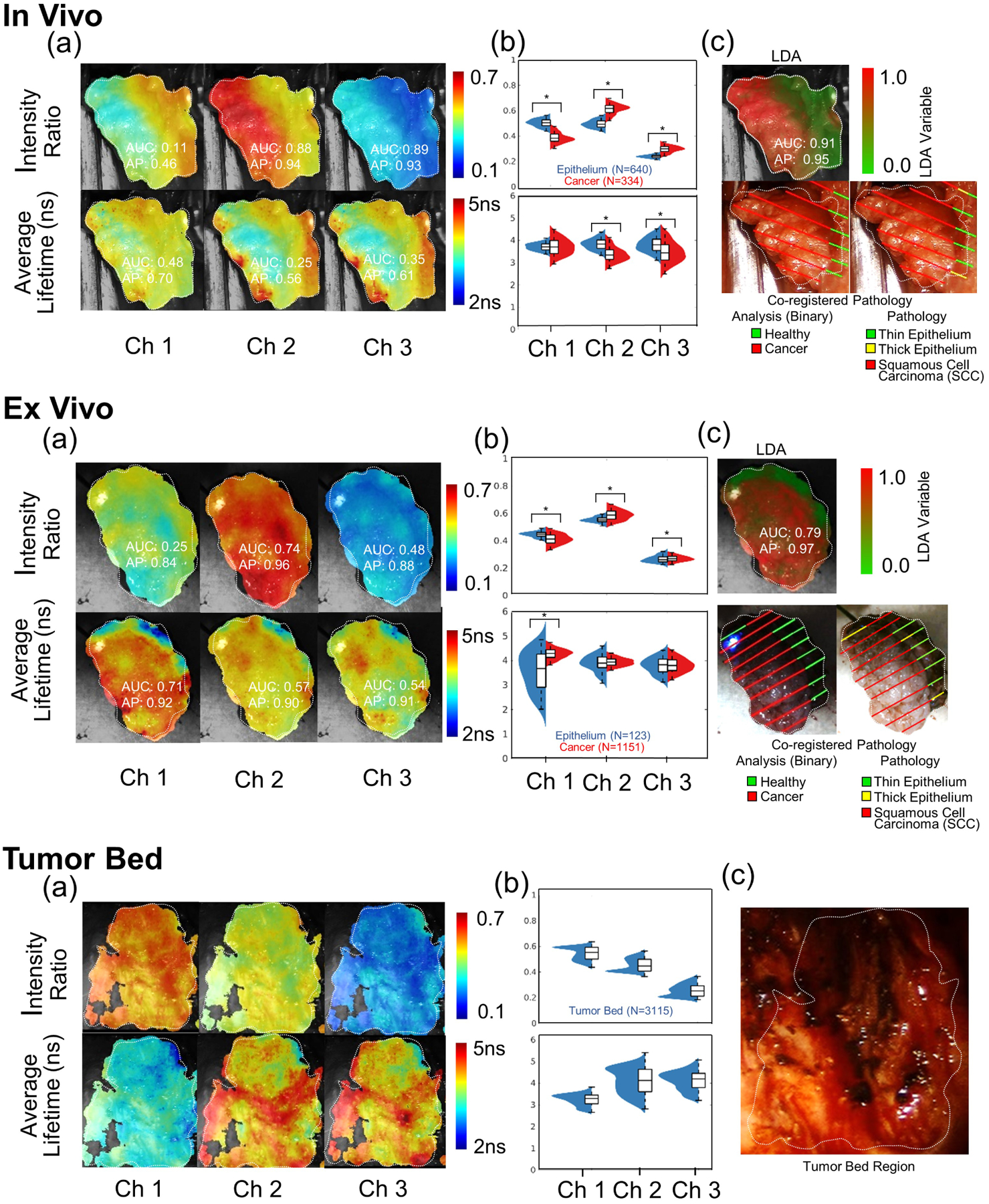
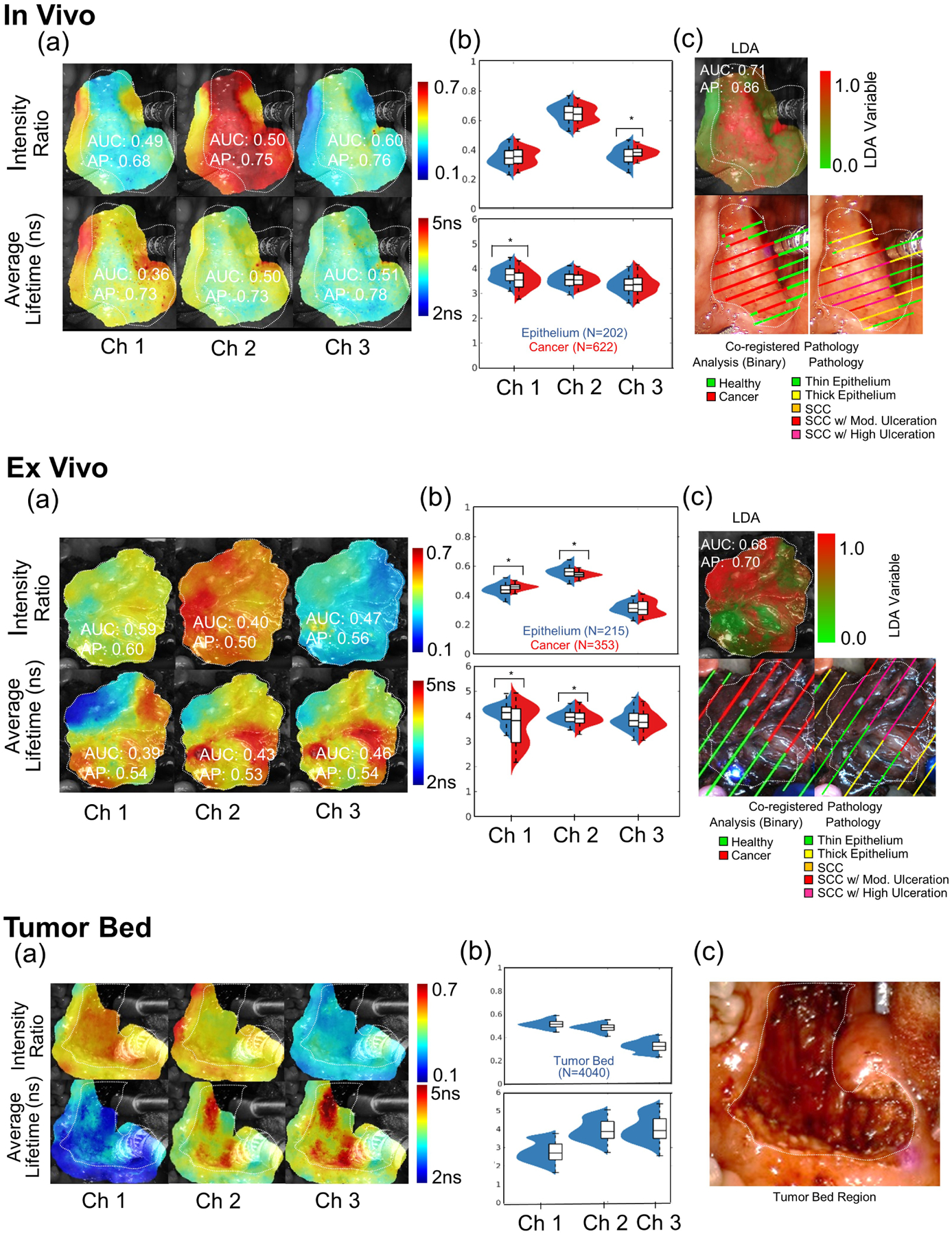
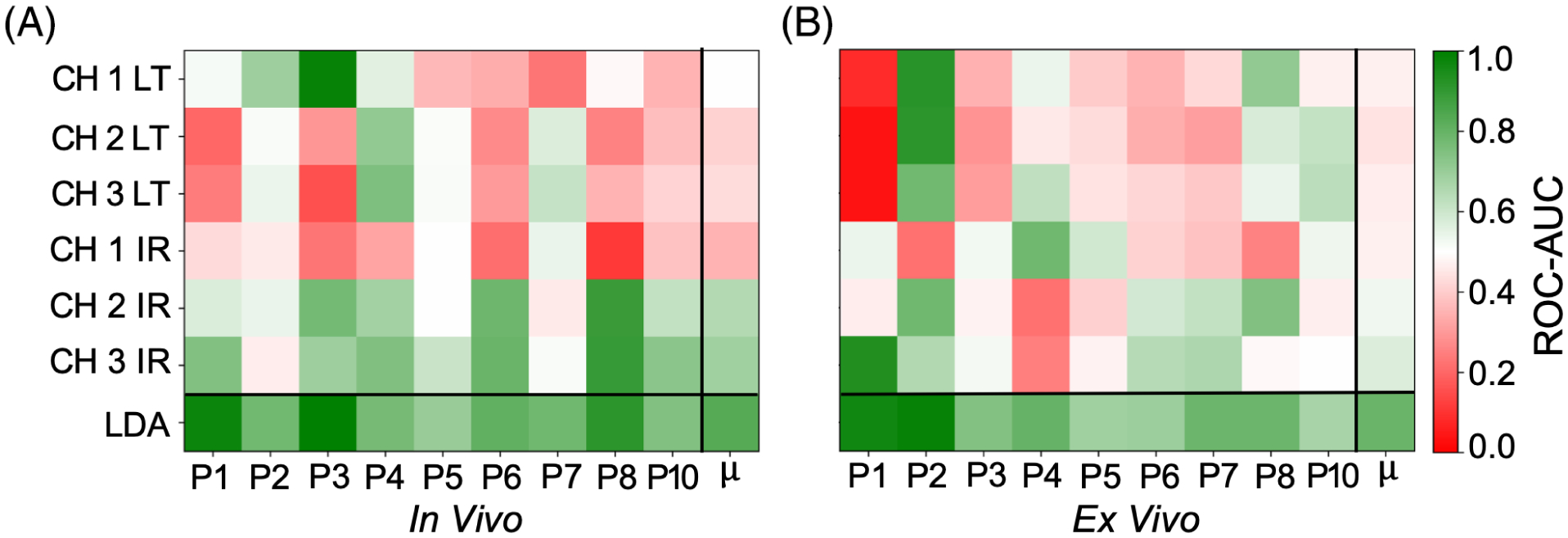
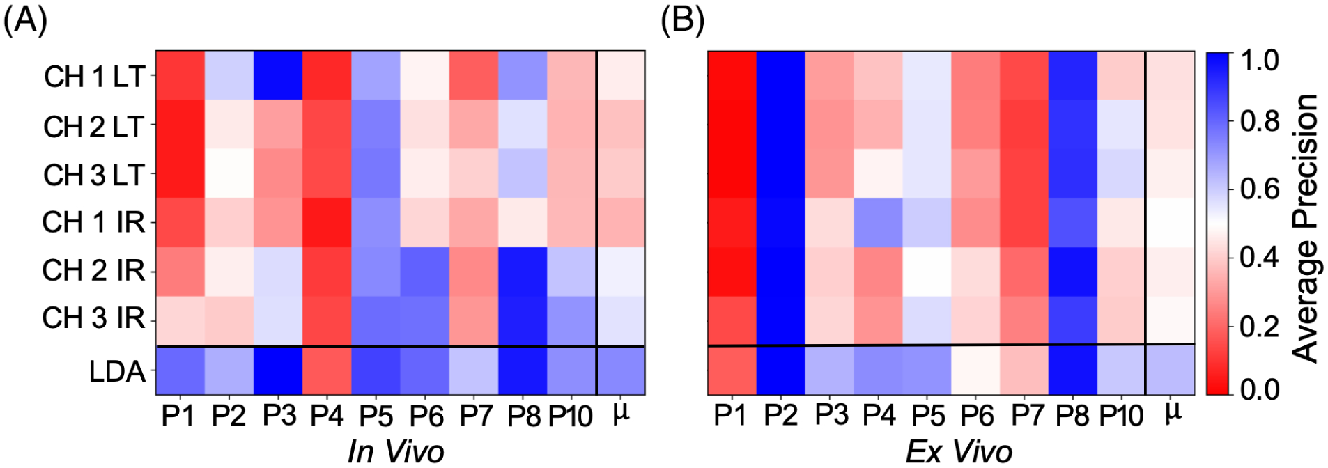
This study evaluates the potential for fluorescence lifetime imaging (FLIm) to enhance intraoperative decisionmaking during robotic-assisted surgery of oropharyngeal cancer. Using a custom built FLIm instrument integrated with the da Vinci robotic surgical platform, we first demonstrate that cancer in epithelial tissue diagnosed by histopathology can be differentiated from surrounding healthy epithelial tissue imaged prior to cancer resection and on the excised specimen. Second, we study the fluorescence properties of tissue imaged at surgical resection margins (tumor bed). Fluorescence lifetimes and spectral intensity ratios were calculated for three spectral channels, producing a set of six FLIm parameters. Current results from 10 patients undergoing TORS procedures demonstrate that healthy epithelium can be resolved from cancer ( < .001) for at least one FLIm parameter. We also showed that a multiparameter linear discriminant analysis approach provides superior discrimination to individual FLIm parameters for tissue imaged both and . Overall, this study highlights the potential for FLIm to be developed into a diagnostic tool for clinical cancer applications of the oropharynx. This technique could help to circumvent the issues posed by the lack of tactile feedback associated with robotic surgical platforms to better enable cancer delineation.
本研究评估了荧光寿命成像(FLIm)在口咽癌机器人辅助手术中增强术中决策的潜力。使用与达芬奇机器人手术平台集成的定制FLIm仪器,我们首先证明,通过组织病理学诊断的上皮组织中的癌症可以与癌症切除前在切除标本上成像的周围健康上皮组织区分开来。其次,我们研究了手术切缘(肿瘤床)成像组织的荧光特性。计算了三个光谱通道的荧光寿命和光谱强度比,产生了一组六个FLIm参数。目前10例接受经口机器人手术(TORS)的患者的结果表明,对于至少一个FLIm参数,健康上皮可以与癌症区分开来(<.001)。我们还表明,多参数线性判别分析方法对成像组织的单个FLIm参数具有更好的判别能力。总体而言,本研究突出了FLIm发展成为口咽临床癌症应用诊断工具的潜力。该技术有助于规避与机器人手术平台相关的缺乏触觉反馈所带来的问题,以更好地实现癌症轮廓的描绘。