,.
Invest Ophthalmol Vis Sci. 2020 Jul 1;61(8):15. doi: 10.1167/iovs.61.8.15.
To describe the pathology of AMD in eyes with geographic atrophy (GA) using confocal scanning laser ophthalmoscopy (SLO) blue light autofluorescence (BAF), and near-infrared (IR) AF and to correlate it with the histology and immunohistochemistry analysis at the margins of the GA lesion.
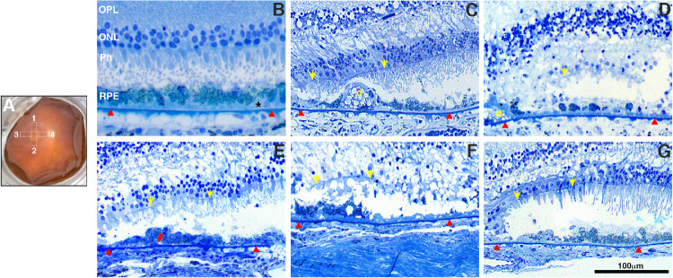
Enucleated, fixed eyes from seventeen donors with GA were imaged and analyzed by BAF-SLO, IRAF-SLO, and by fundus macroscopy (FM). Tissue from the margins of the GA lesions was cut and processed for resin embedding and histology or cryosectioning and fluorescence in the green and far-red channels, and immunohistochemistry to assess markers of inflammation. Isolated DNA from donors was genotyped for single nucleotide polymorphisms (SNPs) previously shown to be risk factors for the development and progression of AMD.
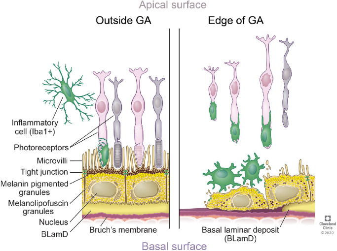
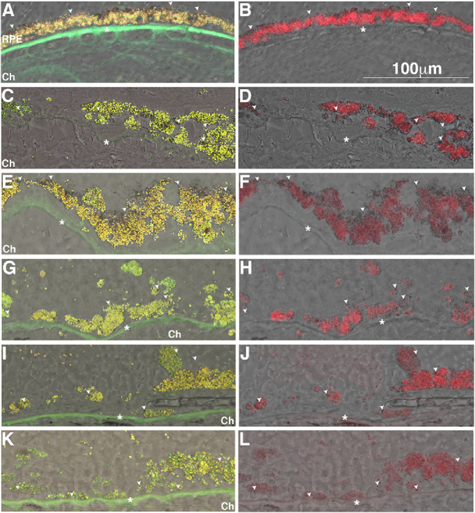
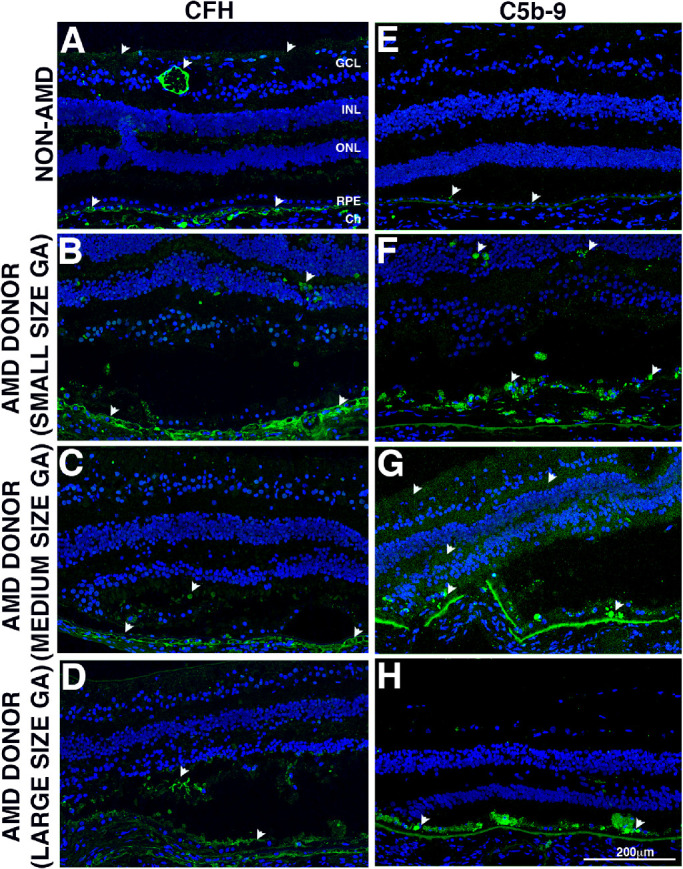
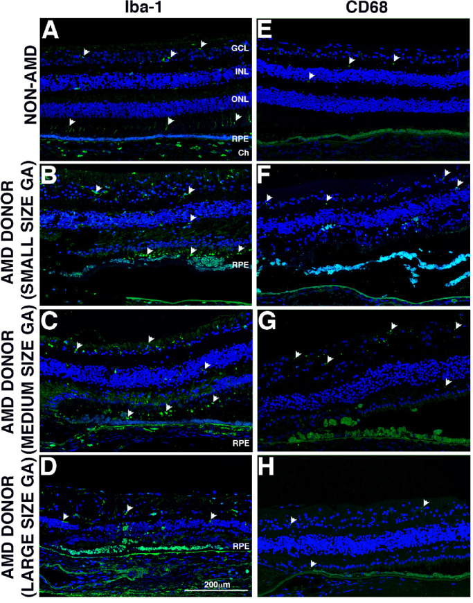
Around the leading edge of the GA lesions we observed hypertrophic RPE cells with cytoplasm filled with granules fluorescent both in the far-red and green-red channels; abundant microglia and macrophage; deposition of complement factor H (CFH) in Bruch's membrane (BM) and increased membrane attack complex (MAC) on RPE cells.
Fluorescence imaging of cryosections of RPE cells around the leading edge of the GA lesions suggest that IRAF-SLO visualizes mostly melanin-related compounds. In addition, medium-size GA atrophy displayed the most significant changes in inflammation markers.
利用共焦扫描激光检眼镜(SLO)蓝光自发荧光(BAF)、近红外(IR)AF 描述伴萎缩性地图状变性(GA)的 AMD 眼部的病理学,并将其与 GA 病变边缘的组织病理学和免疫组织化学分析相关联。
对 17 例 GA 供体眼进行 SLO-BAF、SLO-IRA、眼底宏观照相术(FM)成像和分析。从 GA 病变边缘切取组织,进行树脂包埋和组织学检查,或进行冰冻切片,用绿色通道和远红通道荧光、免疫组织化学检测炎症标志物。从供体中分离的 DNA 进行单核苷酸多态性(SNP)基因分型,这些 SNP 先前被证明是 AMD 发生和进展的危险因素。
在 GA 病变的前缘,我们观察到肥大的 RPE 细胞,其细胞质充满了远红和红绿通道均荧光的颗粒;大量的小胶质细胞和巨噬细胞;Bruch 膜(BM)中补体因子 H(CFH)沉积和 RPE 细胞上的膜攻击复合物(MAC)增加。
GA 病变前缘 RPE 细胞冷冻切片的荧光成像表明,IRA-SLO 主要可视化与黑色素相关的化合物。此外,中大型 GA 萎缩显示出炎症标志物的最显著变化。