Department of Dental Hygiene, College of Health Science, Gachon University, Inchoen 21936, Korea.
Department of Oral Anatomy and Developmental Biology, School of Dentistry, Kyung Hee University, Seoul 02453, Korea.
Int J Environ Res Public Health. 2020 Jul 9;17(14):4956. doi: 10.3390/ijerph17144956.
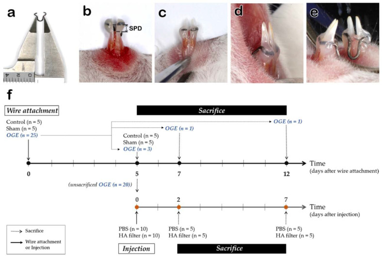
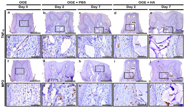
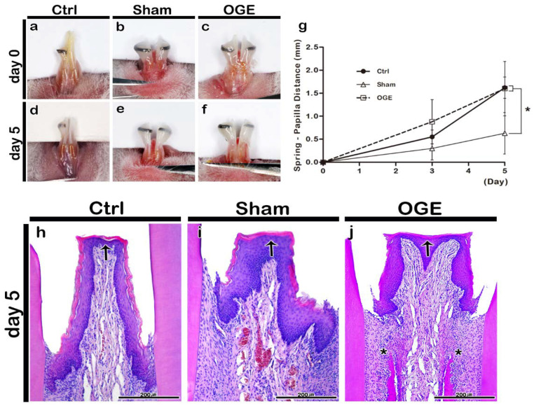
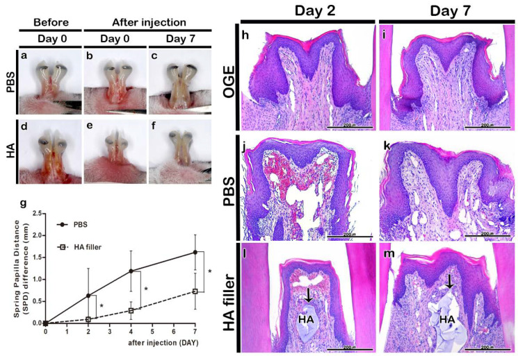
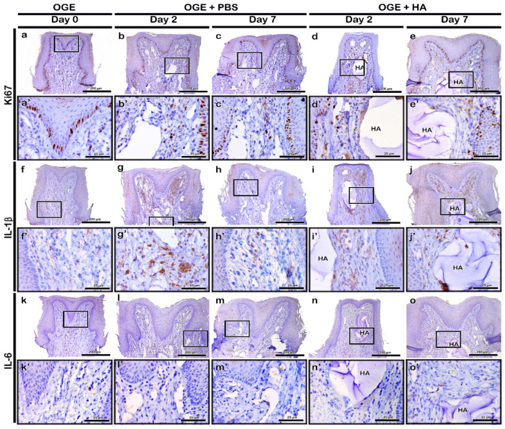
The black triangle resulting from interdental papilla (IDP) loss is associated with poor aesthetics and difficulty in pronunciation and food impaction. There is limited knowledge of gingival tissue inflammatory response to hyaluronic acid (HA) filler injection, a minimally invasive IDP reconstruction method. This study aimed to examine the morphological and histological changes in IDP and the inflammatory cytokine localization to the IDP post-HA filler injection using an open gingival embrasure (OGE) mouse model. Mice from the control, sham, and OGE groups were attached with reference, inactive, and activated wires for 5 days, respectively. The degree of IDP loss was determined based on the spring-papilla distance (SPD). Morphological and histological changes in the OGE group injected with phosphate-buffered saline (PBS) or HA fillers were examined on days 2 and 7 post-injection. Immunohistochemical analysis was performed to determine the localization patterns of tumor necrosis factor (TNF)-α, interleukin (IL)-1β, IL-6, myeloperoxidase (MPO), and Ki67. Five days post-wire attachment, the control and OGE groups exhibited a significantly higher SPD than the sham group ( < 0.0167). The SPD of the HA filler injection group was significantly lower than that of the PBS injection group on days 2, 4, and 7 post-injection ( < 0.05). The IDP of the OGE group was wide and flat. HA filler was stable in the connective tissue underlying the epithelial tissue even on day 7 post-injection. TNF-α, IL-1β, IL-6, MPO, and Ki67 were highly localized to the connective tissue surrounding the filler on day 2, which decreased on day 7 post-injection. Thus, HA filler can safely and successfully reconstruct the IDP in cases of OGE.
牙间乳头(IDP)丧失导致的黑三角与美观度差、发音困难和食物嵌塞有关。对于透明质酸(HA)填充剂注射这种微创 IDP 重建方法,人们对牙龈组织的炎症反应知之甚少。本研究旨在使用开放式龈裂(OGE)小鼠模型,检查 IDP 中 HA 填充剂注射后的形态和组织学变化以及 IDP 中炎症细胞因子的定位。对照组、假手术组和 OGE 组的小鼠分别用参考、不活动和活动的线附着 5 天。根据龈乳头距离(SPD)确定 IDP 丧失的程度。在注射后第 2 和第 7 天,检查 OGE 组注射磷酸盐缓冲盐水(PBS)或 HA 填充剂后的形态和组织学变化。进行免疫组织化学分析以确定肿瘤坏死因子(TNF)-α、白细胞介素(IL)-1β、IL-6、髓过氧化物酶(MPO)和 Ki67 的定位模式。在附着线后 5 天,对照组和 OGE 组的 SPD 明显高于假手术组(<0.0167)。HA 填充剂注射组的 SPD 在注射后第 2、4 和 7 天明显低于 PBS 注射组(<0.05)。OGE 组的 IDP 宽阔而平坦。HA 填充剂在注射后第 7 天仍稳定存在于上皮组织下方的结缔组织中。TNF-α、IL-1β、IL-6、MPO 和 Ki67 在注射后第 2 天高度定位于填充剂周围的结缔组织,在第 7 天减少。因此,HA 填充剂可安全有效地重建 OGE 中的 IDP。