Institute of Dental Sciences, Hebrew University of Jerusalem, Jerusalem, Israel.
Joint Replacement and Reconstructive Surgery Unit, Orthopaedic Surgery Complex, Hadassah Mount Scopus Hospital, Jerusalem, Israel.
Ann Rheum Dis. 2020 Oct;79(10):1370-1380. doi: 10.1136/annrheumdis-2020-217072. Epub 2020 Jul 14.
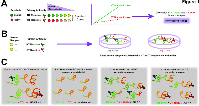
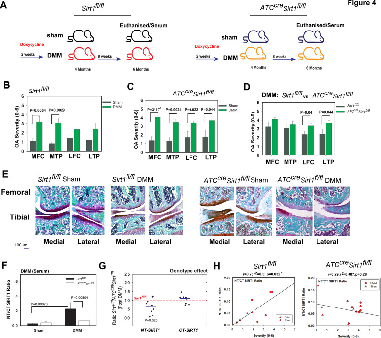
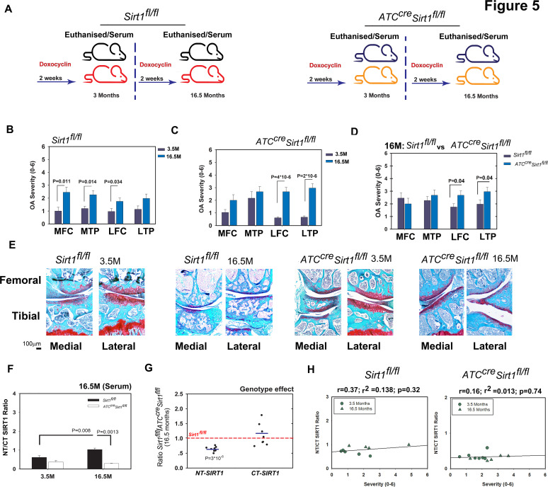
Previous work has established that the deacetylase sirtuin-1 (SIRT1) is cleaved by cathepsin B in chondrocytes subjected to proinflammatory stress, yielding a stable but inactive N-terminal (NT) polypeptide (75SIRT1) and a C-terminal (CT) fragment. The present work examined if chondrocyte-derived NT-SIRT1 is detected in serum and may serve as an investigative and exploratory biomarker of osteoarthritis (OA).
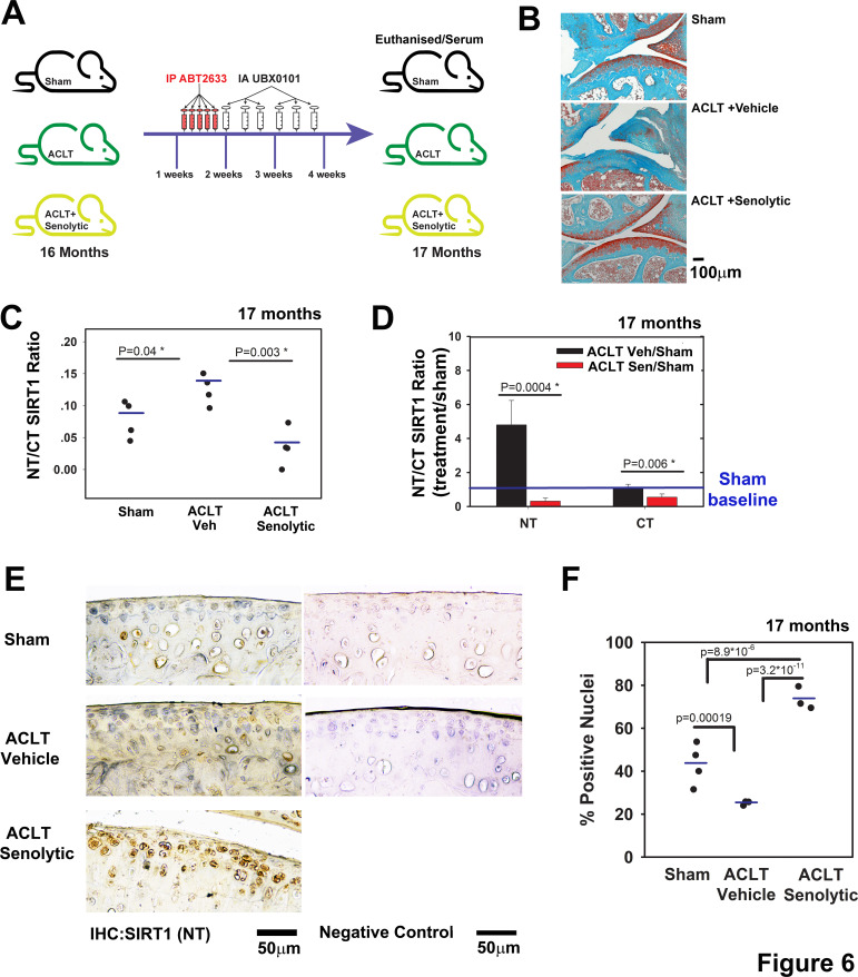
We developed a novel ELISA assay to measure the ratio of NT to CT of SIRT1 in the serum of human individuals and mice subjected to post-traumatic OA (PTOA) or age-dependent OA (ADOA). We additionally monitored NT/CT SIRT1 in mice subject to ADOA/PTOA followed by senolytic clearance. Human chondrosenescent and non-senescent chondrocytes were exposed to cytokines and analysed for apoptosis and NT/CT SIRT1 ratio in conditioned medium.
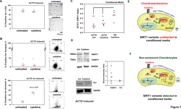
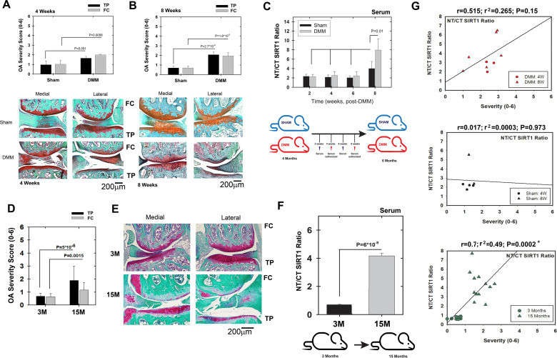
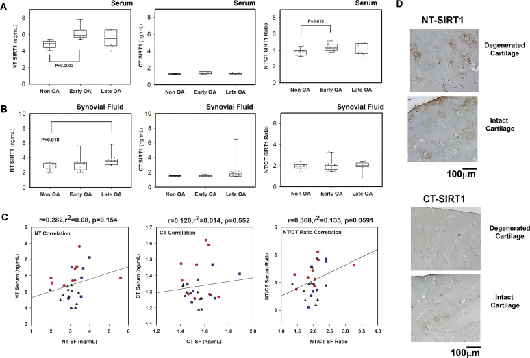
Wild-type mice with PTOA or ADOA of moderate severity exhibited increased serum NT/CT SIRT1 ratio. In contrast, this ratio remained low in cartilage-specific knockout mice despite similar or increased PTOA and ADOA severity. Local clearance of senescent chondrocytes from old mice with post-traumatic injury resulted in a lower NT/CT ratio and reduced OA severity. While primary chondrocytes exhibited NT/CT ratio increased in conditioned media after prolonged cytokine stimulation, this increase was not evident in cytokine-stimulated chondrosenescent cells. Finally, serum NT/CT ratio was elevated in humans with early-stage OA.
Increased levels of serum NT/CT SIRT1 ratio correlated with moderate OA in both mice and humans, stemming at least in part from non-senescent chondrocyte apoptosis, possibly a result of prolonged inflammatory insult.
先前的研究已经证实,软骨细胞在受到促炎应激时,组织蛋白酶 B 会将去乙酰化酶 Sirtuin-1(SIRT1)切割成稳定但无活性的 N 端(NT)多肽(75SIRT1)和 C 端(CT)片段。本研究旨在探讨软骨细胞来源的 NT-SIRT1 是否存在于血清中,并可能作为骨关节炎(OA)的研究和探索性生物标志物。
我们开发了一种新的 ELISA 检测方法,用于测量人血清中和经创伤后 OA(PTOA)或年龄相关性 OA(ADOA)处理的小鼠血清中 SIRT1 的 NT 与 CT 的比值。我们还监测了接受 ADOA/PTOA 治疗后进行衰老细胞清除的小鼠的 NT/CT SIRT1。将软骨衰老细胞和非衰老细胞暴露于细胞因子中,分析条件培养基中的细胞凋亡和 NT/CT SIRT1 比值。
具有中等严重程度 PTOA 或 ADOA 的野生型小鼠表现出升高的血清 NT/CT SIRT1 比值。相比之下,尽管 PTOA 和 ADOA 严重程度相似或增加,软骨特异性 基因敲除小鼠中的该比值仍保持较低水平。对创伤后老年小鼠进行衰老细胞局部清除可导致较低的 NT/CT 比值和减轻 OA 严重程度。虽然原代软骨细胞在长时间细胞因子刺激后培养基中的 NT/CT 比值增加,但在细胞因子刺激的软骨衰老细胞中则没有明显增加。最后,血清 NT/CT 比值在早期 OA 患者中升高。
血清 NT/CT SIRT1 比值的升高与小鼠和人类的中度 OA 相关,至少部分源自非衰老软骨细胞凋亡,可能是长期炎症损伤的结果。