Department of Joint Surgery and Sports Medicine, Zhongnan Hospital of Wuhan University, Wuhan, 430071, China.
J Orthop Surg Res. 2020 Jul 15;15(1):264. doi: 10.1186/s13018-020-01786-4.
Aseptic necrosis of the femoral head (ANFH) has a high incidence in the community and causes substantial problems with health as well as economic and social stress. Core decompression is the most commonly used treatment for early ANFH. Although many studies have reported on the efficacy of femoral head core decompression surgery for ANFH, there are still some shortcomings in assessing the severity of femoral head necrosis, the location distribution, and changes in necrotic lesions before and after surgery. Magnetic resonance imaging (MRI) and equivalent sphere model analysis were used to further clarify the clinical efficacy of percutaneous multiple small-diameter drilling core decompression in patients with ANFH.
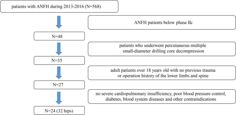
From July 2013 to November 2016, 24 patients (32 cases of the hip joint) with ANFH who underwent percutaneous multiple small-diameter drilling core decompression were selected, and a retrospective analysis was conducted. MRI as well as VAS, OHS-C, and HHS scores were used to evaluate joint function in all patients before and 6, 12, and 24 months after the operation.
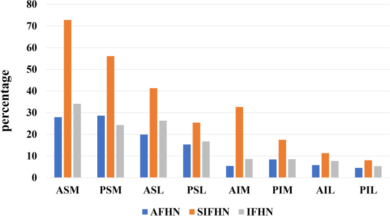
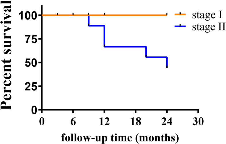
Twenty-four months after the operation, 10 hips were amputated. The survival rates of alcoholic femoral head necrosis (AFNH), idiopathic femoral head necrosis (IFHN), and steroid-induced femoral head necrosis (SIFHN) patients at 24 months were 100%, 85.7% (- 2 hips), and 0.0% (- 8 hips), respectively. The MRI and equivalent sphere analysis results revealed that the anterior superior medial quadrant was the area most prone to osteonecrosis, and the posterior superior medial quadrant was the area second most prone to necrosis. After the operation, the average percentage of the AFHN necrosis area in the total volume of the femoral head decreased from 14.5 to 10.3%, and the average percentage of the IFHN necrosis area decreased from 16.3 to 9.2%; however, the average percentage of the necrosis area for SIFHN increased from 30.4 to 33.1%.
Percutaneous multiple small-diameter drilling core decompression significantly reduced the lesion volume for AFHN and IFHN, but the effect on SIFHN was not good.
股骨头无菌性坏死(ANFH)在社区中发病率较高,不仅对健康造成严重影响,还给患者带来经济和社会压力。核心减压是治疗早期 ANFH 的最常用方法。虽然许多研究报告了股骨头核心减压手术治疗 ANFH 的疗效,但在评估股骨头坏死的严重程度、坏死病变的位置分布以及手术前后的变化方面仍存在一些不足。磁共振成像(MRI)和等效球体模型分析进一步阐明了经皮多枚小直径钻孔核心减压治疗 ANFH 的临床疗效。
回顾性分析 2013 年 7 月至 2016 年 11 月收治的 24 例(32 髋)ANFH 患者,均行经皮多枚小直径钻孔核心减压术,所有患者术前及术后 6、12、24 个月均采用 VAS、OHS-C、HHS 评分评估关节功能。
术后 24 个月,10 髋截肢。酒精性股骨头坏死(AFNH)、特发性股骨头坏死(IFHN)和激素性股骨头坏死(SIFHN)患者的 24 个月生存率分别为 100%(-2 髋)、85.7%(-2 髋)和 0.0%(-8 髋)。MRI 和等效球体分析结果显示,前上内侧象限是最易发生骨坏死的区域,其次是后上内侧象限。术后,AFNH 坏死面积占股骨头总体积的百分比从 14.5%降至 10.3%,IFHN 坏死面积百分比从 16.3%降至 9.2%;而 SIFHN 的坏死面积百分比从 30.4%增加到 33.1%。
经皮多枚小直径钻孔核心减压术可显著减少 AFHN 和 IFHN 的病变体积,但对 SIFHN 的效果不佳。