Wooff Yvette, Cioanca Adrian V, Chu-Tan Joshua A, Aggio-Bruce Riemke, Schumann Ulrike, Natoli Riccardo
The John Curtin School of Medical Research, The Australian National University, Canberra, ACT, Australia.
The ANU Medical School, The Australian National University, Canberra, ACT, Australia.
Front Cell Neurosci. 2020 Jun 25;14:160. doi: 10.3389/fncel.2020.00160. eCollection 2020.
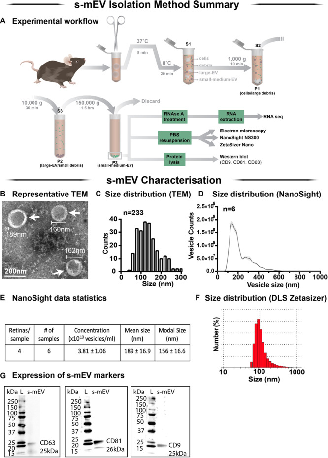
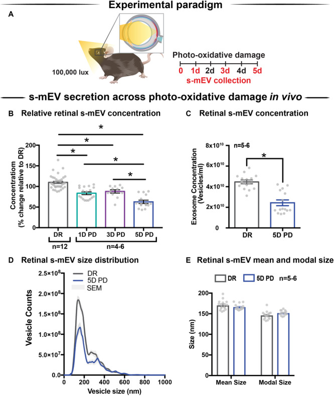
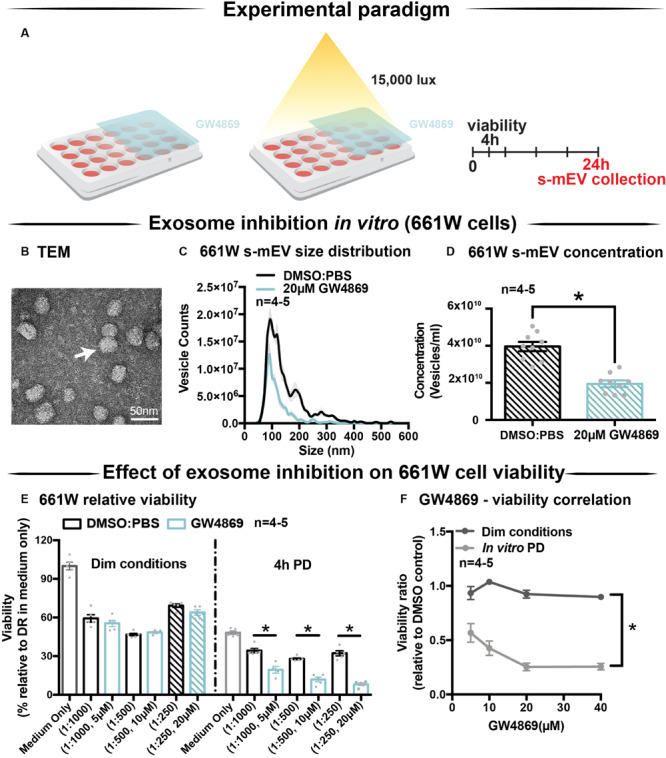
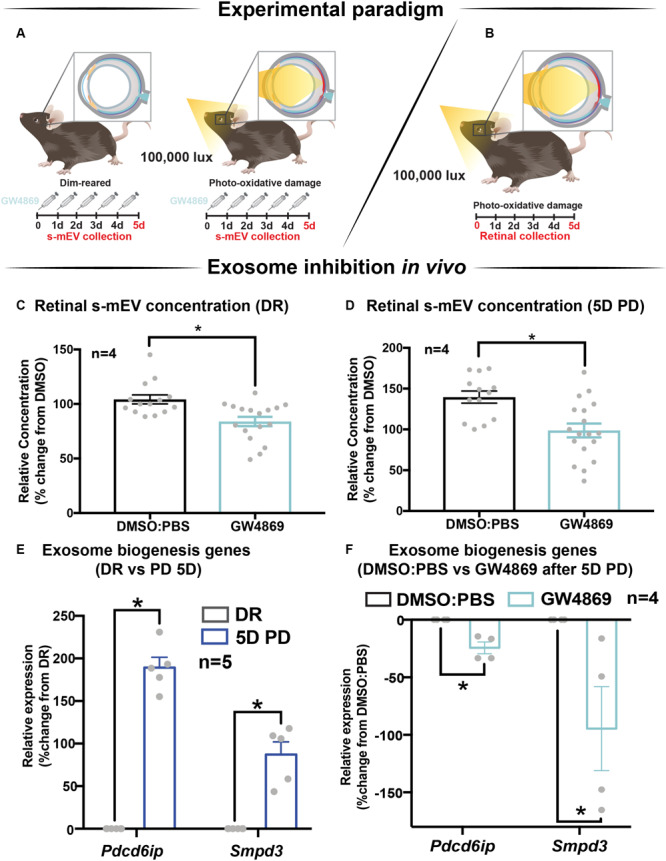
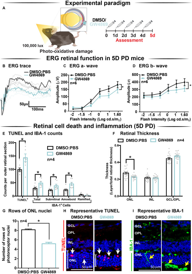
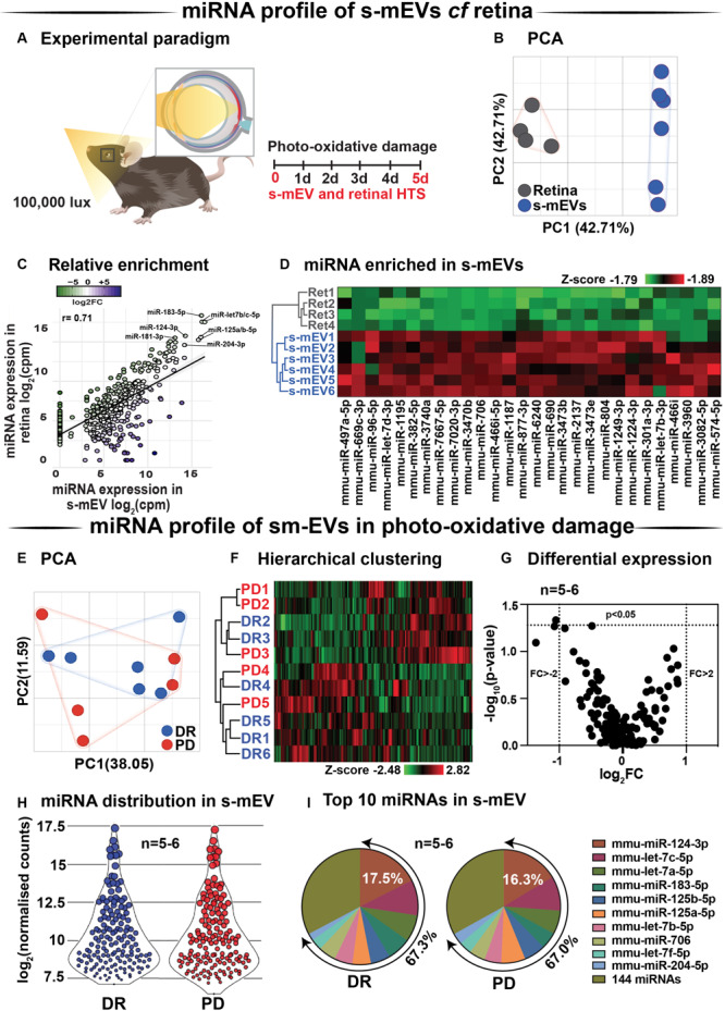
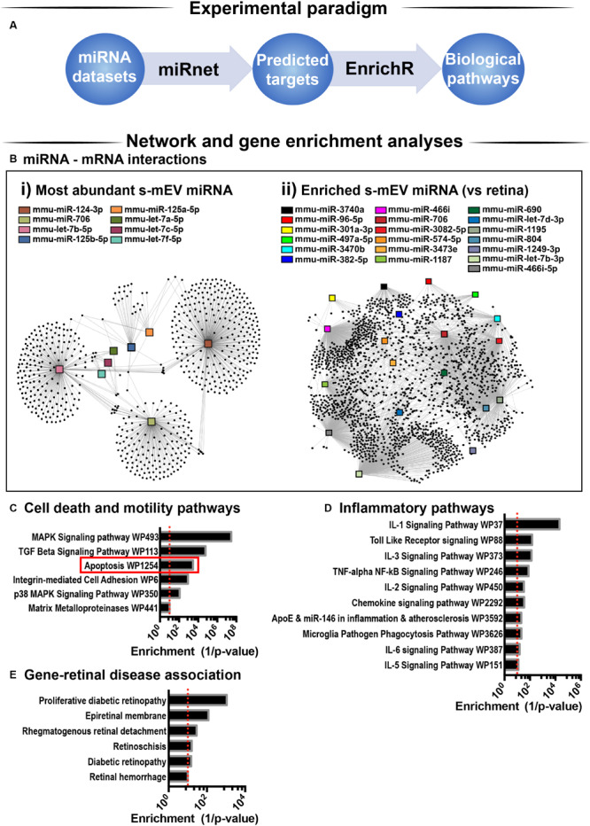
Photoreceptor cell death and inflammation are known to occur progressively in retinal degenerative diseases such as age-related macular degeneration (AMD). However, the molecular mechanisms underlying these biological processes are largely unknown. Extracellular vesicles (EV) are essential mediators of cell-to-cell communication with emerging roles in the modulation of immune responses. EVs, including exosomes, encapsulate and transfer microRNA (miRNA) to recipient cells and in this way can modulate the environment of recipient cells. Dysregulation of EVs however is correlated to a loss of cellular homeostasis and increased inflammation. In this work we investigated the role of isolated retinal small-medium sized EV (s-mEV) which includes exosomes in both the healthy and degenerating retina. Isolated s-mEV from normal retinas were characterized using dynamic light scattering, transmission electron microscopy and western blotting, and quantified across 5 days of photo-oxidative damage-induced degeneration using nanotracking analysis. Small RNAseq was used to characterize the miRNA cargo of retinal s-mEV isolated from healthy and damaged retinas. Finally, the effect of exosome inhibition on cell-to-cell miRNA transfer and immune modulation was conducted using systemic daily administration of exosome inhibitor GW4869 and hybridization of s-mEV-abundant miRNA, miR-124-3p. Electroretinography and immunohistochemistry was performed to assess functional and morphological changes to the retina as a result of GW4869-induced exosome depletion. Results demonstrated an inverse correlation between s-mEV concentration and photoreceptor survivability, with a decrease in s-mEV numbers following degeneration. Small RNAseq revealed that s-mEVs contained uniquely enriched miRNAs in comparison to in whole retinal tissue, however, there was no differential change in the s-mEV miRNAnome following photo-oxidative damage. Exosome inhibition via the use of GW4869 was also found to exacerbate retinal degeneration, with reduced retinal function and increased levels of inflammation and cell death demonstrated following photo-oxidative damage in exosome-inhibited mice. Further, GW4869-treated mice displayed impaired translocation of photoreceptor-derived miR-124-3p to the inner retina during damage. Taken together, we propose that retinal s-mEV and their miRNA cargo play an essential role in maintaining retinal homeostasis through immune-modulation, and have the potential to be used in targeted gene therapy for retinal degenerative diseases.
已知在年龄相关性黄斑变性(AMD)等视网膜退行性疾病中,光感受器细胞死亡和炎症会逐渐发生。然而,这些生物学过程背后的分子机制在很大程度上尚不清楚。细胞外囊泡(EV)是细胞间通讯的重要介质,在免疫反应调节中发挥着新作用。包括外泌体在内的EV会包裹并将微小RNA(miRNA)转移至受体细胞,从而调节受体细胞的环境。然而,EV失调与细胞内稳态丧失和炎症增加相关。在这项研究中,我们调查了分离出的视网膜中小尺寸EV(s-mEV,包括外泌体)在健康视网膜和退化视网膜中的作用。使用动态光散射、透射电子显微镜和蛋白质印迹法对从正常视网膜中分离出的s-mEV进行表征,并通过纳米追踪分析对光氧化损伤诱导的退化5天内的s-mEV进行定量。使用小RNA测序来表征从健康和受损视网膜中分离出的视网膜s-mEV的miRNA含量。最后,通过每天全身给予外泌体抑制剂GW4869以及对富含s-mEV的miRNA miR-124-3p进行杂交,研究外泌体抑制对细胞间miRNA转移和免疫调节的影响。进行视网膜电图和免疫组织化学检查,以评估由于GW4869诱导的外泌体耗竭而导致的视网膜功能和形态变化。结果表明,s-mEV浓度与光感受器存活率呈负相关,退化后s-mEV数量减少。小RNA测序显示,与整个视网膜组织相比,s-mEV含有独特富集的miRNA,然而,光氧化损伤后s-mEV的miRNA组没有差异变化。还发现通过使用GW4869抑制外泌体会加剧视网膜退化,在外泌体抑制的小鼠中,光氧化损伤后视网膜功能降低,炎症和细胞死亡水平增加。此外,GW4869处理的小鼠在损伤期间光感受器来源的miR-124-3p向内视网膜的转运受损。综上所述,我们认为视网膜s-mEV及其miRNA含量通过免疫调节在维持视网膜内稳态中起重要作用,并且有潜力用于视网膜退行性疾病的靶向基因治疗。