Department of Clinical Neurological Sciences, Division of Neurosurgery, Western University, London, Ontario, Canada.
Imaging Research Laboratories, Robarts Research Institute Canada, Western University, London, Ontario, Canada.
Hum Brain Mapp. 2020 Nov;41(16):4500-4517. doi: 10.1002/hbm.25137. Epub 2020 Jul 17.
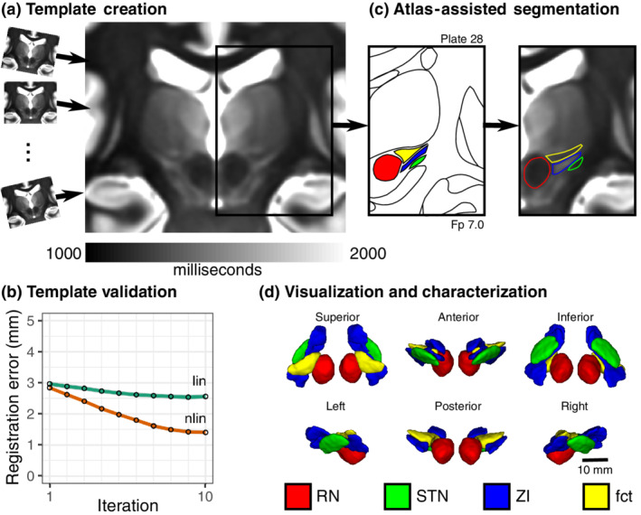
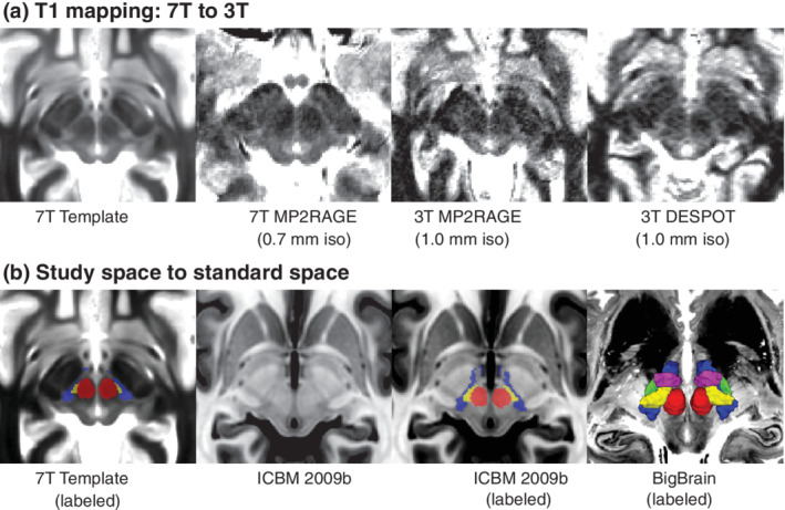
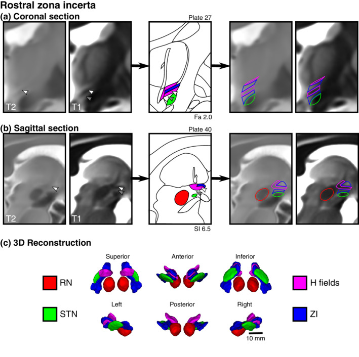
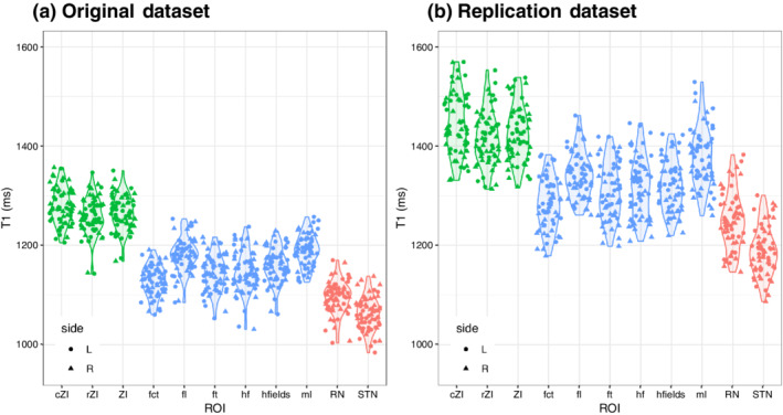
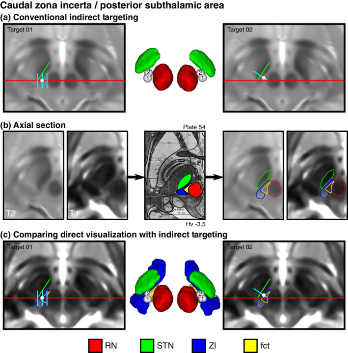
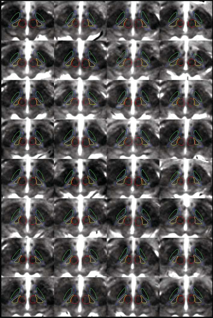
The zona incerta (ZI) is a small gray matter region of the deep brain first identified in the 19th century, yet direct in vivo visualization and characterization has remained elusive. Noninvasive detection of the ZI and surrounding region could be critical to further our understanding of this widely connected but poorly understood deep brain region and could contribute to the development and optimization of neuromodulatory therapies. We demonstrate that high resolution (submillimetric) longitudinal (T1) relaxometry measurements at high magnetic field strength (7 T) can be used to delineate the ZI from surrounding white matter structures, specifically the fasciculus cerebellothalamicus, fields of Forel (fasciculus lenticularis, fasciculus thalamicus, and field H), and medial lemniscus. Using this approach, we successfully derived in vivo estimates of the size, shape, location, and tissue characteristics of substructures in the ZI region, confirming observations only previously possible through histological evaluation that this region is not just a space between structures but contains distinct morphological entities that should be considered separately. Our findings pave the way for increasingly detailed in vivo study and provide a structural foundation for precise functional and neuromodulatory investigation.
神经内分泌核(ZI)是大脑深部的一个小的灰质区域,于 19 世纪首次被发现,但直接的体内可视化和特征描述仍然难以实现。ZI 及其周围区域的非侵入性检测对于进一步了解这个广泛连接但了解甚少的深部脑区可能至关重要,并有助于神经调节治疗的发展和优化。我们证明,在高磁场强度(7T)下进行高分辨率(亚毫米)纵向(T1)弛豫测量可用于从周围的白质结构(特别是小脑丘脑束、Forel 区(豆状核束、丘脑束和 H 区)和内侧丘系)中描绘 ZI。使用这种方法,我们成功地推导出了 ZI 区域内亚结构的大小、形状、位置和组织特征的体内估计值,证实了只有通过组织学评估才能观察到的发现,即该区域不仅是结构之间的空间,还包含应单独考虑的不同形态实体。我们的研究结果为越来越详细的体内研究铺平了道路,并为精确的功能和神经调节研究提供了结构基础。