Xu Yanwu
Neurosurgery Department, People's Hospital of Lanling County, Linyi, Shandong province, 277700, China.
World J Surg Oncol. 2020 Jul 16;18(1):169. doi: 10.1186/s12957-020-01949-x.
Malignant brain tumors have been a serious threat to human health worldwide. This study aims to investigate the role of miR-136-3p in glioma development.
Hematoxylin-eosin staining (H&E) staining was used to determine the pathologic alterations of glioma tissues. Quantitative real-time PCR (qRT-PCR) analysis and GEO2R analysis was performed to examine the expression of miRNAs and genes. Western blot was applied to detect the protein expression. Cell counting kit-8 (CCK-8) and colony formation were used to analyze the glioma cell growth. Trans-well assay was used to determine the cell migration. Annexin V-FITC/PI staining was conducted to determine the cell apoptosis of transfected glioma cells. The dual-luciferase reporter assay was carried out to confirm the binding sites of miR-136-3p on 3' untranslated regions (3' UTR) of Kruppel-like factor 7 (KLF7). Tumor-bearing experiment in nude mice was performed to comprehensively investigate the role of miR-136-3p/KLF7 axis in gliomas.
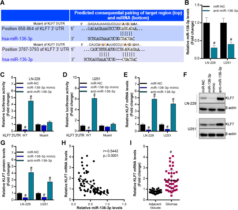
Firstly, the results showed that miR-136-3p was decreased in glioma tissues compared with adjacent tissues. Overexpression of miR-136-3p significantly inhibited cell growth of LN-229 and U251 by decreasing expression of Cyclin A1 and PCNA (proliferating cell nuclear antigen), and it suppressed glioma cell migration by downregulating N-cadherin and elevating E-cadherin levels, and it also promotes glioma cell apoptosis by promoting Bcl2-associated X (Bax) expression but suppressing Bcl-2 expression. Furthermore, we observed that KLF7 was a direct target of miR-136-3p, and KLF7 was negatively regulated by miR-136-3p in glioma cells. Finally, overexpression of KLF7 partly blocked miR-136-3p-induced inhibition of tumor growth in vitro and in vivo.
Targeting miR-136-3p/KLF7 axis might be a novel manner to counter against gliomas.
恶性脑肿瘤一直是全球范围内对人类健康的严重威胁。本研究旨在探讨miR-136-3p在胶质瘤发生发展中的作用。
采用苏木精-伊红染色(H&E)确定胶质瘤组织的病理改变。进行定量实时聚合酶链反应(qRT-PCR)分析和GEO2R分析以检测微小RNA(miRNA)和基因的表达。应用蛋白质免疫印迹法检测蛋白质表达。使用细胞计数试剂盒-8(CCK-8)和集落形成实验分析胶质瘤细胞的生长情况。采用Transwell实验检测细胞迁移。进行膜联蛋白V-异硫氰酸荧光素/碘化丙啶(Annexin V-FITC/PI)染色以确定转染胶质瘤细胞的凋亡情况。进行双荧光素酶报告基因实验以确认miR-136-3p在Kruppel样因子7(KLF7)的3'非翻译区(3'UTR)上的结合位点。在裸鼠中进行荷瘤实验以全面研究miR-136-3p/KLF7轴在胶质瘤中的作用。
首先,结果显示与相邻组织相比,miR-136-3p在胶质瘤组织中表达降低。miR-136-3p的过表达通过降低细胞周期蛋白A1和增殖细胞核抗原(PCNA)的表达显著抑制LN-229和U251细胞的生长,通过下调N-钙黏蛋白并提高E-钙黏蛋白水平抑制胶质瘤细胞迁移,并且通过促进Bcl-2相关X蛋白(Bax)表达但抑制Bcl-2表达促进胶质瘤细胞凋亡。此外,我们观察到KLF7是miR-136-3p的直接靶点,并且在胶质瘤细胞中KLF7受到miR-136-3p的负调控。最后,KLF7的过表达部分阻断了miR-136-3p在体外和体内诱导的肿瘤生长抑制作用。
靶向miR-136-3p/KLF7轴可能是对抗胶质瘤的一种新方法。