Department of Otorhinolaryngology, University Hospital Ulm, Frauensteige 12, 89075 Ulm, Germany.
Department of Internal Medicine III, University of Ulm, Helmholtzstr. 10, 89081 Ulm, Germany.
Int J Mol Sci. 2020 Jul 22;21(15):5181. doi: 10.3390/ijms21155181.
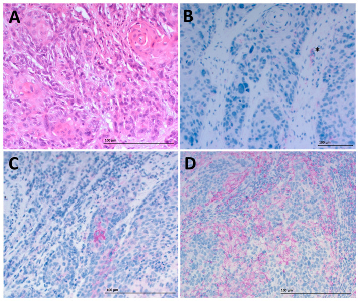
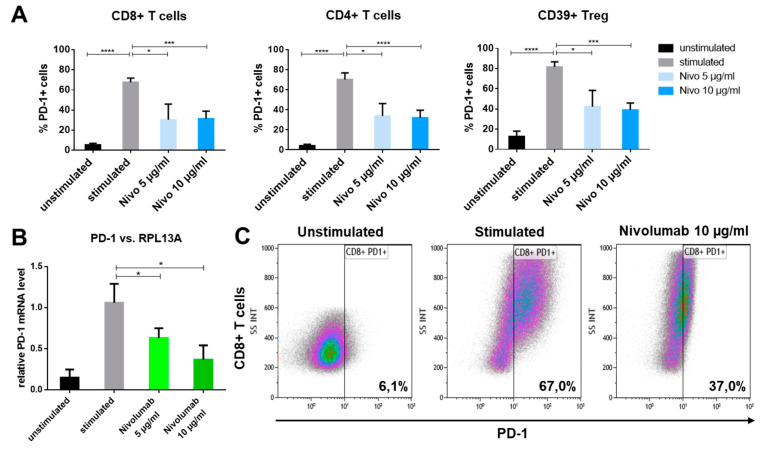
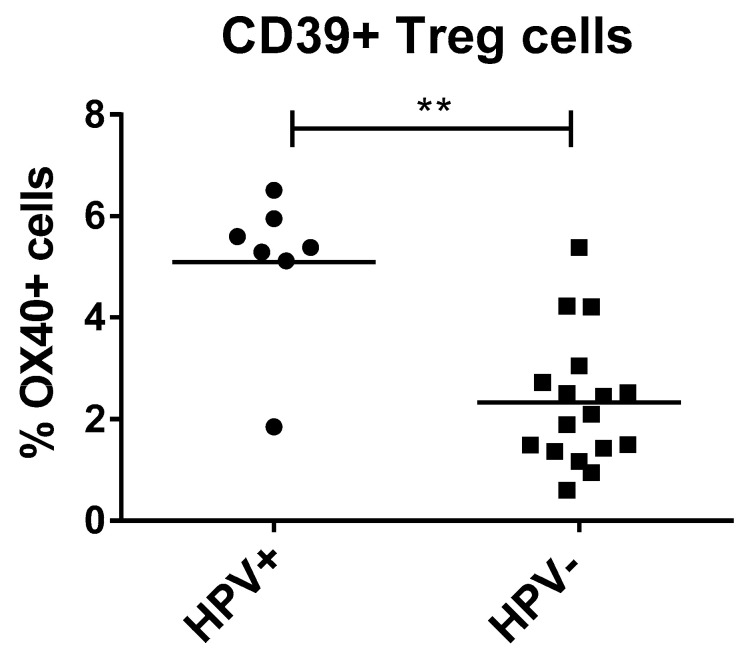
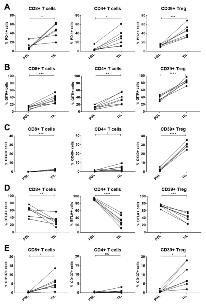
Endogenous control mechanisms, including immune checkpoints and immunosuppressive cells, are exploited in the process of tumorigenesis to weaken the anti-tumor immune response. Cancer treatment by chemotherapy or immune checkpoint inhibition can lead to changes of checkpoint expression, which influences therapy success. Peripheral blood lymphocytes (PBL) and tumor-infiltrating lymphocytes (TIL) were isolated from head and neck squamous cell carcinoma (HNSCC) patients ( = 23) and compared to healthy donors ( = 23). Immune checkpoint expression (programmed cell death ligand 1 (PD-1), tumor necrosis factor receptor (TNFR)-related (GITR), CD137, tumor necrosis factor receptor superfamily member 4 (TNFRSF4) (OX40), t-cell immunoglobulin and mucin-domain containing-3 (TIM3), B- and T-lymphocyte attenuator (BTLA), lymphocyte-activation gene 3 (LAG3)) was determined on immune cells by flow cytometry. PD-L1 expression was detected on tumor tissue by immunohistochemistry. Immune cells were treated with immuno- and chemotherapeutics to investigate treatment-specific change in immune checkpoint expression, in vitro. Specific changes of immune checkpoint expression were identified on PBL and TIL of HNSCC patients compared to healthy donors. Various chemotherapeutics acted differently on the expression of immune checkpoints. Changes of checkpoint expression were significantly less pronounced on regulatory T cells compared to other lymphocyte populations. Nivolumab treatment significantly reduced the receptor PD-1 on all analyzed T cell populations, in vitro. The specific immune checkpoint expression patterns in HNSCC patients and the investigated effects of immunomodulatory agents may improve the development and efficacy of targeted immunotherapy.
内源性控制机制,包括免疫检查点和免疫抑制细胞,在肿瘤发生过程中被利用来削弱抗肿瘤免疫反应。化疗或免疫检查点抑制的癌症治疗可导致检查点表达的变化,这影响治疗的成功。从头颈部鳞状细胞癌(HNSCC)患者(n = 23)和健康供体(n = 23)中分离外周血淋巴细胞(PBL)和肿瘤浸润淋巴细胞(TIL)。通过流式细胞术测定免疫细胞上的免疫检查点表达(程序性细胞死亡配体 1(PD-1)、肿瘤坏死因子受体(TNFR)相关(GITR)、CD137、肿瘤坏死因子受体超家族成员 4(TNFRSF4)(OX40)、T 细胞免疫球蛋白和粘蛋白结构域包含-3(TIM3)、B 和 T 淋巴细胞衰减器(BTLA)、淋巴细胞激活基因 3(LAG3))。通过免疫组织化学法检测肿瘤组织中 PD-L1 的表达。用免疫和化疗药物处理免疫细胞,以研究体外治疗特异性的免疫检查点表达变化。与健康供体相比,HNSCC 患者的 PBL 和 TIL 上识别出免疫检查点表达的特定变化。各种化疗药物对免疫检查点的表达有不同的作用。与其他淋巴细胞群相比,调节性 T 细胞的检查点表达变化不那么明显。在体外,nivolumab 治疗显著降低了所有分析的 T 细胞群上的受体 PD-1。HNSCC 患者的特定免疫检查点表达模式和所研究的免疫调节剂的作用可能会改善靶向免疫治疗的开发和疗效。