Li Jia, Li Yanchun, Atakan Muhammed M, Kuang Jujiao, Hu Yang, Bishop David J, Yan Xu
College of Physical Education, Southwest University, Chongqing 400715, China.
Institute for Health and Sport (iHeS), Victoria University, P.O. Box 14428, Melbourne 8001, Australia.
Antioxidants (Basel). 2020 Jul 24;9(8):656. doi: 10.3390/antiox9080656.
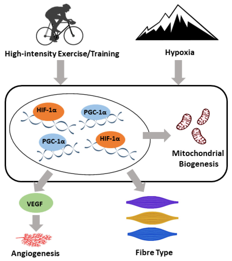
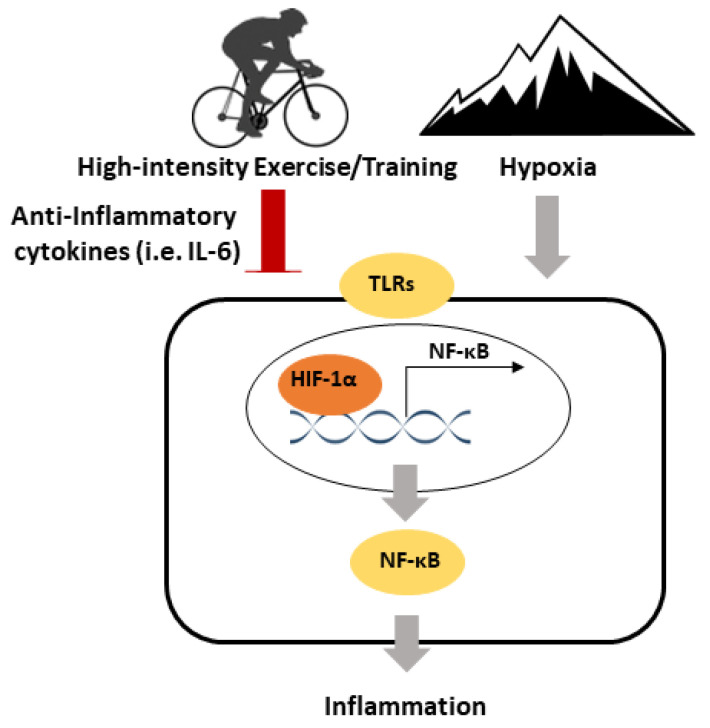
High-intensity exercise/training, especially interval exercise/training, has gained popularity in recent years. Hypoxic training was introduced to elite athletes half a century ago and has recently been adopted by the general public. In the current review, we have summarised the molecular adaptive responses of skeletal muscle to high-intensity exercise/training, focusing on mitochondrial biogenesis, angiogenesis, and muscle fibre composition. The literature suggests that (peroxisome proliferator-activated receptor gamma coactivator 1-alpha) PGC-1α, vascular endothelial growth factor (VEGF), and hypoxia-inducible factor 1-alpha (HIF1-α) might be the main mediators of skeletal muscle adaptations to high-intensity exercises in hypoxia. Exercise is known to be anti-inflammatory, while the effects of hypoxia on inflammatory signalling are more complex. The anti-inflammatory effects of a single session of exercise might result from the release of anti-inflammatory myokines and other cytokines, as well as the downregulation of Toll-like receptor signalling, while training-induced anti-inflammatory effects may be due to reductions in abdominal and visceral fat (which are main sources of pro-inflammatory cytokines). Hypoxia can lead to inflammation, and inflammation can result in tissue hypoxia. However, the hypoxic factor HIF1-α is essential for preventing excessive inflammation. Disease-induced hypoxia is related to an upregulation of inflammatory signalling, but the effects of exercise-induced hypoxia on inflammation are less conclusive. The effects of high-intensity exercise under hypoxia on skeletal muscle molecular adaptations and inflammatory signalling have not been fully explored and are worth investigating in future studies. Understanding these effects will lead to a more comprehensive scientific basis for maximising the benefits of high-intensity exercise.
近年来,高强度运动/训练,尤其是间歇运动/训练越来越受欢迎。低氧训练在半个世纪前被引入精英运动员群体,最近已被普通大众所采用。在本综述中,我们总结了骨骼肌对高强度运动/训练的分子适应性反应,重点关注线粒体生物发生、血管生成和肌纤维组成。文献表明,过氧化物酶体增殖物激活受体γ共激活因子1α(PGC-1α)、血管内皮生长因子(VEGF)和低氧诱导因子1α(HIF1-α)可能是骨骼肌在低氧环境下对高强度运动产生适应性变化的主要介质。众所周知,运动具有抗炎作用,而低氧对炎症信号传导的影响则更为复杂。单次运动的抗炎作用可能源于抗炎性肌动蛋白和其他细胞因子的释放,以及Toll样受体信号传导的下调,而训练诱导的抗炎作用可能是由于腹部和内脏脂肪(促炎细胞因子的主要来源)减少所致。低氧可导致炎症,而炎症又可导致组织低氧。然而,低氧因子HIF1-α对于预防过度炎症至关重要。疾病诱导的低氧与炎症信号传导上调有关,但运动诱导的低氧对炎症的影响尚无定论。低氧环境下高强度运动对骨骼肌分子适应性和炎症信号传导的影响尚未得到充分研究,值得未来深入探讨。了解这些影响将为最大化高强度运动的益处提供更全面的科学依据。