Department of Ophthalmology, University of Bonn, Bonn, Germany.
GRADE Reading Center, Bonn, Germany.
Invest Ophthalmol Vis Sci. 2020 Aug 3;61(10):19. doi: 10.1167/iovs.61.10.19.
To examine longitudinal changes of retinal thickness and retinal sensitivity in patients with intermediate age-related macular degeneration (iAMD) and predominantly reticular pseudodrusen (RPD).
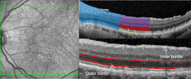
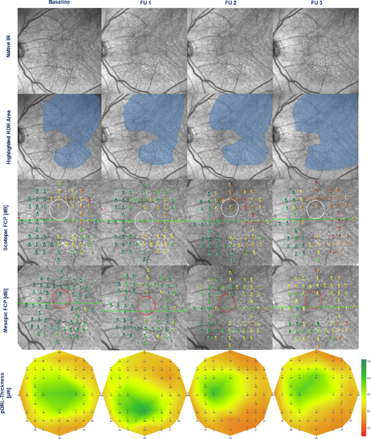
At baseline 30 eyes of 25 iAMD patients underwent optical coherence tomography imaging, mesopic and scotopic fundus-controlled perimetry (FCP) with follow-up examinations at month 12 (20 eyes), 24 (12 eyes), and 36 (11 eyes). Thicknesses of different retinal layers and results of FCP testing (n = 56 stimuli) were spatially and longitudinally analyzed using linear mixed-effects models.
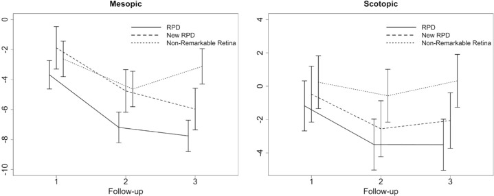
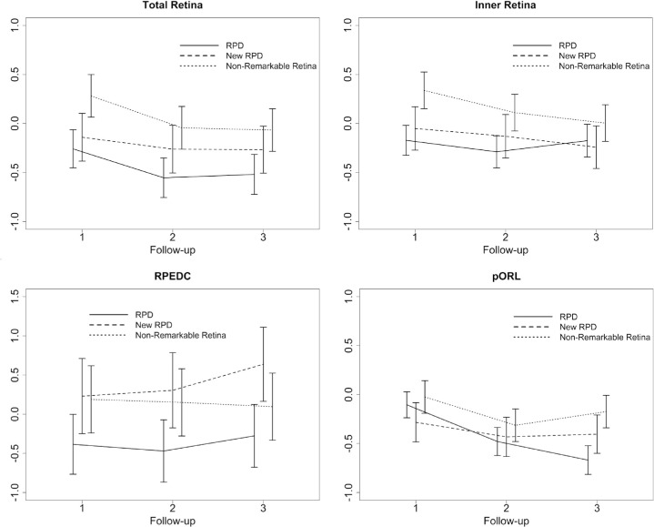
At baseline, the thickness of the partial outer retinal layer (pORL, 70.21 vs. 77.47 µm) and both mesopic (16.60 vs. 18.72 dB) and scotopic (12.14 vs. 18.67 dB) retinal sensitivity were decreased in areas with RPD compared with unremarkable areas (P < 0.001). Over three years, mean change of pORL was -0.66 normative standard deviation (SD; i.e., z-score, P < 0.001) for regions with existing RPD, -0.40 SD (P < 0.001) for regions with new occurring RPD, and -0.17 SD (P = 0.041) in unremarkable regions. Decrease of scotopic and mesopic sensitivity over three years was more pronounced in areas with existing (-3.51 and -7.76 dB) and new occurring RPD (-2.06 and -5.97 dB). Structure-function analysis revealed that 1 SD decrease of pORL thickness was associated with a sensitivity reduction of 3.47 dB in scotopic and 0.79 dB in mesopic testing.
This study demonstrates progressive outer retinal degeneration and impairment of photoreceptor function in eyes with iAMD and RPD over three years. Preservation of outer retinal thickness and reduction of RPD formation may constitute meaningful surrogate endpoints in interventional trials on eyes with AMD and RPD aiming to slow outer retinal degeneration.
研究中间型年龄相关性黄斑变性(iAMD)和主要为网状假性玻璃膜疣(RPD)患者的视网膜厚度和视网膜敏感性的纵向变化。
在基线时,25 名 iAMD 患者的 30 只眼接受了光学相干断层扫描成像、中值和暗视眼底控制视野检查(FCP),并在第 12 个月(20 只眼)、24 个月(12 只眼)和 36 个月(11 只眼)进行了随访检查。使用线性混合效应模型对不同视网膜层的厚度和 FCP 测试结果(n = 56 个刺激)进行空间和纵向分析。
在基线时,与无异常区域相比,RPD 区域的部分外层视网膜厚度(pORL,70.21 µm 比 77.47 µm)和中值(16.60 dB 比 18.72 dB)和暗视(12.14 dB 比 18.67 dB)视网膜敏感性均降低(P < 0.001)。在三年内,对于存在 RPD 的区域,pORL 的平均变化为-0.66 个正常标准差(SD;即 z 分数,P < 0.001),对于出现新的 RPD 的区域为-0.40 SD(P < 0.001),在无异常区域为-0.17 SD(P = 0.041)。在三年内,对于存在 RPD(-3.51 和-7.76 dB)和新出现的 RPD(-2.06 和-5.97 dB)的区域,暗视和中值敏感性的下降更为明显。结构-功能分析表明,pORL 厚度减少 1 SD 与暗视和中值测试中的敏感性降低 3.47 dB 和 0.79 dB 相关。
本研究表明,在三年内,iAMD 和 RPD 患者的外视网膜逐渐退化和光感受器功能受损。保留外视网膜厚度和减少 RPD 形成可能成为旨在减缓外视网膜退化的 AMD 和 RPD 患者干预试验的有意义的替代终点。