Liu Jing, Li Linlin, Yang Yang, Hong Bei, Chen Xi, Xie Qiwei, Han Hua
National Laboratory of Pattern Recognition, Institute of Automation, Chinese Academy of Sciences, Beijing, China.
School of Artificial Intelligence, School of Future Technology, University of Chinese Academy of Sciences, Beijing, China.
Front Neurosci. 2020 Jul 21;14:599. doi: 10.3389/fnins.2020.00599. eCollection 2020.
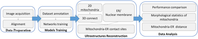
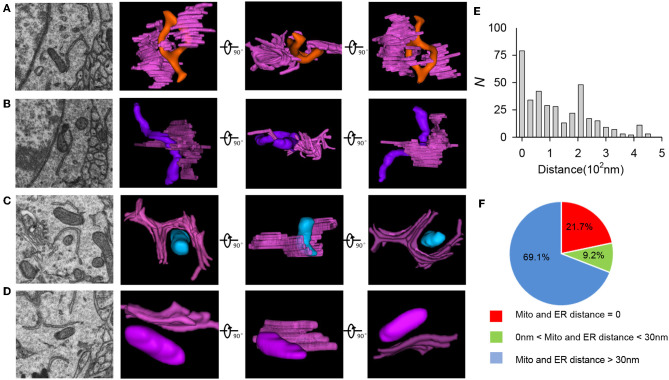
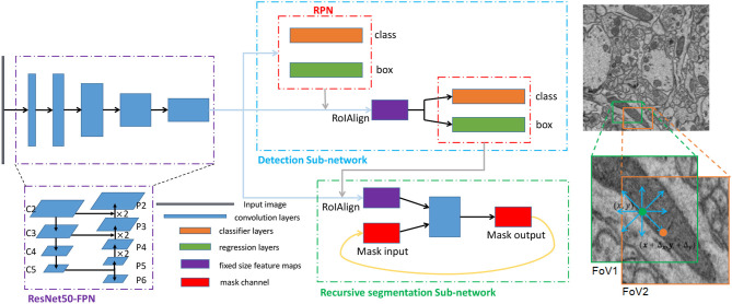
Together, mitochondria and the endoplasmic reticulum (ER) occupy more than 20% of a cell's volume, and morphological abnormality may lead to cellular function disorders. With the rapid development of large-scale electron microscopy (EM), manual contouring and three-dimensional (3D) reconstruction of these organelles has previously been accomplished in biological studies. However, manual segmentation of mitochondria and ER from EM images is time consuming and thus unable to meet the demands of large data analysis. Here, we propose an automated pipeline for mitochondrial and ER reconstruction, including the mitochondrial and ER contact sites (MAMs). We propose a novel recurrent neural network to detect and segment mitochondria and a fully residual convolutional network to reconstruct the ER. Based on the sparse distribution of synapses, we use mitochondrial context information to rectify the local misleading results and obtain 3D mitochondrial reconstructions. The experimental results demonstrate that the proposed method achieves state-of-the-art performance.
线粒体和内质网(ER)加起来占据了细胞体积的20%以上,形态异常可能导致细胞功能紊乱。随着大规模电子显微镜(EM)的迅速发展,此前在生物学研究中已经完成了对这些细胞器的手动轮廓描绘和三维(3D)重建。然而,从EM图像中手动分割线粒体和内质网非常耗时,因此无法满足大数据分析的需求。在这里,我们提出了一种用于线粒体和内质网重建的自动化流程,包括线粒体-内质网接触位点(MAMs)。我们提出了一种新颖的循环神经网络来检测和分割线粒体,并使用全残差卷积网络来重建内质网。基于突触的稀疏分布,我们利用线粒体上下文信息来纠正局部误导性结果,从而获得3D线粒体重建。实验结果表明,所提出的方法达到了当前最优的性能。