• 文献检索
  • 文档翻译
  • 深度研究
  • 学术资讯
  • Suppr Zotero 插件Zotero 插件
  • 邀请有礼
  • 套餐&价格
  • 历史记录
应用&插件
Suppr Zotero 插件Zotero 插件浏览器插件Mac 客户端Windows 客户端微信小程序
定价
高级版会员购买积分包购买API积分包
服务
文献检索文档翻译深度研究API 文档MCP 服务
关于我们
关于 Suppr公司介绍联系我们用户协议隐私条款
关注我们

Suppr 超能文献

核心技术专利:CN118964589B侵权必究
粤ICP备2023148730 号-1Suppr @ 2026

文献检索

告别复杂PubMed语法,用中文像聊天一样搜索,搜遍4000万医学文献。AI智能推荐,让科研检索更轻松。

立即免费搜索

文件翻译

保留排版,准确专业,支持PDF/Word/PPT等文件格式,支持 12+语言互译。

免费翻译文档

深度研究

AI帮你快速写综述,25分钟生成高质量综述,智能提取关键信息,辅助科研写作。

立即免费体验

使用3D监督卷积网络对电子显微镜数据进行自动线粒体分割

Automatic Mitochondria Segmentation for EM Data Using a 3D Supervised Convolutional Network.

作者信息

Xiao Chi, Chen Xi, Li Weifu, Li Linlin, Wang Lu, Xie Qiwei, Han Hua

机构信息

Institute of Automation, Chinese Academy of Sciences, Beijing, China.

School of Future Technology, University of Chinese Academy of Sciences, Beijing, China.

出版信息

Front Neuroanat. 2018 Nov 2;12:92. doi: 10.3389/fnana.2018.00092. eCollection 2018.

DOI:10.3389/fnana.2018.00092
PMID:30450040
原文链接:https://pmc.ncbi.nlm.nih.gov/articles/PMC6224513/
Abstract

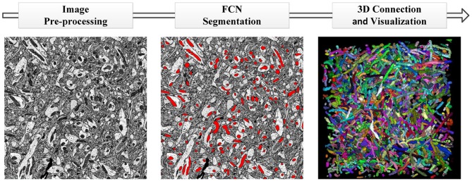
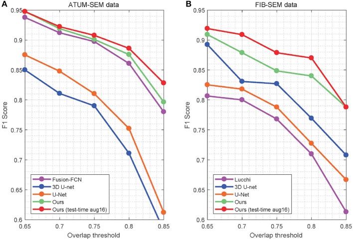
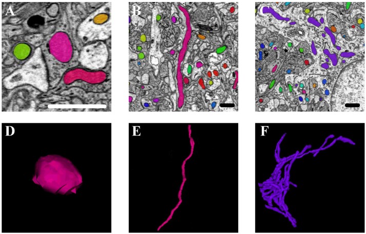
Recent studies have supported the relation between mitochondrial functions and degenerative disorders related to ageing, such as Alzheimer's and Parkinson's diseases. Since these studies have exposed the need for detailed and high-resolution analysis of physical alterations in mitochondria, it is necessary to be able to perform segmentation and 3D reconstruction of mitochondria. However, due to the variety of mitochondrial structures, automated mitochondria segmentation and reconstruction in electron microscopy (EM) images have proven to be a difficult and challenging task. This paper puts forward an effective and automated pipeline based on deep learning to realize mitochondria segmentation in different EM images. The proposed pipeline consists of three parts: (1) utilizing image registration and histogram equalization as image pre-processing steps to maintain the consistency of the dataset; (2) proposing an effective approach for 3D mitochondria segmentation based on a volumetric, residual convolutional and deeply supervised network; and (3) employing a 3D connection method to obtain the relationship of mitochondria and displaying the 3D reconstruction results. To our knowledge, we are the first researchers to utilize a 3D fully residual convolutional network with a deeply supervised strategy to improve the accuracy of mitochondria segmentation. The experimental results on anisotropic and isotropic EM volumes demonstrate the effectiveness of our method, and the Jaccard index of our segmentation (91.8% in anisotropy, 90.0% in isotropy) and F1 score of detection (92.2% in anisotropy, 90.9% in isotropy) suggest that our approach achieved state-of-the-art results. Our fully automated pipeline contributes to the development of neuroscience by providing neurologists with a rapid approach for obtaining rich mitochondria statistics and helping them elucidate the mechanism and function of mitochondria.

摘要

最近的研究证实了线粒体功能与衰老相关的退行性疾病(如阿尔茨海默病和帕金森病)之间的关系。由于这些研究揭示了对线粒体物理变化进行详细和高分辨率分析的必要性,因此有必要能够对线粒体进行分割和三维重建。然而,由于线粒体结构的多样性,在电子显微镜(EM)图像中实现线粒体的自动分割和重建已被证明是一项困难且具有挑战性的任务。本文提出了一种基于深度学习的有效且自动化的流程,以实现不同EM图像中的线粒体分割。所提出的流程包括三个部分:(1)利用图像配准和直方图均衡化作为图像预处理步骤,以保持数据集的一致性;(2)基于体积、残差卷积和深度监督网络提出一种有效的三维线粒体分割方法;(3)采用三维连接方法来获取线粒体之间的关系并展示三维重建结果。据我们所知,我们是首批利用具有深度监督策略的三维全残差卷积网络来提高线粒体分割准确性的研究人员。在各向异性和各向同性EM体积上的实验结果证明了我们方法的有效性,我们分割的杰卡德指数(各向异性中为91.8%,各向同性中为90.0%)和检测的F1分数(各向异性中为92.2%,各向同性中为90.9%)表明我们的方法取得了领先的结果。我们的全自动流程为神经科学家提供了一种快速获取丰富线粒体统计数据的方法,并帮助他们阐明线粒体的机制和功能,从而推动了神经科学的发展。

https://cdn.ncbi.nlm.nih.gov/pmc/blobs/c60f/6224513/1a3d70f6da3d/fnana-12-00092-g0009.jpg
https://cdn.ncbi.nlm.nih.gov/pmc/blobs/c60f/6224513/f9093f7272c7/fnana-12-00092-g0001.jpg
https://cdn.ncbi.nlm.nih.gov/pmc/blobs/c60f/6224513/66e3366c4003/fnana-12-00092-g0002.jpg
https://cdn.ncbi.nlm.nih.gov/pmc/blobs/c60f/6224513/cf6105171317/fnana-12-00092-g0003.jpg
https://cdn.ncbi.nlm.nih.gov/pmc/blobs/c60f/6224513/ee396c09a078/fnana-12-00092-g0004.jpg
https://cdn.ncbi.nlm.nih.gov/pmc/blobs/c60f/6224513/741ea0d75fe8/fnana-12-00092-g0005.jpg
https://cdn.ncbi.nlm.nih.gov/pmc/blobs/c60f/6224513/8103168dc914/fnana-12-00092-g0006.jpg
https://cdn.ncbi.nlm.nih.gov/pmc/blobs/c60f/6224513/b090da4989b4/fnana-12-00092-g0007.jpg
https://cdn.ncbi.nlm.nih.gov/pmc/blobs/c60f/6224513/bd4b9e8801ed/fnana-12-00092-g0008.jpg
https://cdn.ncbi.nlm.nih.gov/pmc/blobs/c60f/6224513/1a3d70f6da3d/fnana-12-00092-g0009.jpg

相似文献

1
Automatic Mitochondria Segmentation for EM Data Using a 3D Supervised Convolutional Network.使用3D监督卷积网络对电子显微镜数据进行自动线粒体分割
Front Neuroanat. 2018 Nov 2;12:92. doi: 10.3389/fnana.2018.00092. eCollection 2018.
2
Effective automated pipeline for 3D reconstruction of synapses based on deep learning.基于深度学习的有效自动化突触 3D 重建流水线。
BMC Bioinformatics. 2018 Jul 13;19(1):263. doi: 10.1186/s12859-018-2232-0.
3
Automatic Reconstruction of Mitochondria and Endoplasmic Reticulum in Electron Microscopy Volumes by Deep Learning.通过深度学习对电子显微镜图像中的线粒体和内质网进行自动重建
Front Neurosci. 2020 Jul 21;14:599. doi: 10.3389/fnins.2020.00599. eCollection 2020.
4
3D deeply supervised network for automated segmentation of volumetric medical images.三维深度监督网络在容积医学图像自动分割中的应用。
Med Image Anal. 2017 Oct;41:40-54. doi: 10.1016/j.media.2017.05.001. Epub 2017 May 8.
5
A Novel 3D Connection Algorithm of Mitochondria From ATUM-SEM Stacks Based on Segmentation Information in Context.一种基于上下文分割信息的来自ATUM-SEM堆栈的线粒体新型三维连接算法。
Annu Int Conf IEEE Eng Med Biol Soc. 2018 Jul;2018:5105-5108. doi: 10.1109/EMBC.2018.8513488.
6
HIVE-Net: Centerline-aware hierarchical view-ensemble convolutional network for mitochondria segmentation in EM images.HIVE-Net:用于 EM 图像中线粒体分割的中心感知层次视图集成卷积网络。
Comput Methods Programs Biomed. 2021 Mar;200:105925. doi: 10.1016/j.cmpb.2020.105925. Epub 2021 Jan 10.
7
Automatic segmentation of mitochondria and endolysosomes in volumetric electron microscopy data.在体积电子显微镜数据中对线粒体和内溶酶体进行自动分割。
Comput Biol Med. 2020 Apr;119:103693. doi: 10.1016/j.compbiomed.2020.103693. Epub 2020 Mar 3.
8
UNI-EM: An Environment for Deep Neural Network-Based Automated Segmentation of Neuronal Electron Microscopic Images.UNI-EM:一种基于深度神经网络的神经元电子显微镜图像自动分割环境。
Sci Rep. 2019 Dec 19;9(1):19413. doi: 10.1038/s41598-019-55431-0.
9
Automatic bladder segmentation from CT images using deep CNN and 3D fully connected CRF-RNN.利用深度卷积神经网络和 3D 全连接条件随机场循环神经网络自动进行 CT 图像的膀胱分割。
Int J Comput Assist Radiol Surg. 2018 Jul;13(7):967-975. doi: 10.1007/s11548-018-1733-7. Epub 2018 Mar 19.
10
Semi Supervised Learning with Deep Embedded Clustering for Image Classification and Segmentation.用于图像分类和分割的深度嵌入聚类半监督学习
IEEE Access. 2019;7:11093-11104. doi: 10.1109/ACCESS.2019.2891970. Epub 2019 Jan 9.

引用本文的文献

1
DeepSCEM: A User-Friendly Solution for Deep Learning-Based Image Segmentation in Cellular Electron Microscopy.DeepSCEM:细胞电子显微镜中基于深度学习的图像分割的用户友好解决方案。
Biol Cell. 2025 Sep;117(9):e70032. doi: 10.1111/boc.70032.
2
Morphological multiparameter filtration and persistent homology in mitochondrial image analysis.线粒体图像分析中的形态多参数过滤和持久同调。
PLoS One. 2024 Sep 20;19(9):e0310157. doi: 10.1371/journal.pone.0310157. eCollection 2024.
3
Systems mapping of bidirectional endosomal transport through the crowded cell.

本文引用的文献

1
Large scale three-dimensional reconstruction of an entire Caenorhabditis elegans larva using AutoCUTS-SEM.使用 AutoCUTS-SEM 对整个秀丽隐杆线虫幼虫进行大规模三维重建。
J Struct Biol. 2017 Nov;200(2):87-96. doi: 10.1016/j.jsb.2017.09.010. Epub 2017 Oct 1.
2
An automated pipeline for mitochondrial segmentation on ATUM-SEM stacks.一种用于在ATUM-SEM堆栈上进行线粒体分割的自动化流程。
J Bioinform Comput Biol. 2017 Jun;15(3):1750015. doi: 10.1142/S0219720017500159. Epub 2017 May 26.
3
3D deeply supervised network for automated segmentation of volumetric medical images.
双向内体运输在拥挤细胞中的系统映射。
Curr Biol. 2024 Oct 7;34(19):4476-4494.e11. doi: 10.1016/j.cub.2024.08.026. Epub 2024 Sep 13.
4
A Damaging COL6A3 Variant Alters the MIR31HG-Regulated Response of Chondrocytes in Neocartilage Organoids to Hyperphysiologic Mechanical Loading.一种具有破坏性的 COL6A3 变体改变了软骨细胞在类软骨器官中对超生理机械加载的 MIR31HG 调节反应。
Adv Sci (Weinh). 2024 Sep;11(36):e2400720. doi: 10.1002/advs.202400720. Epub 2024 Jul 17.
5
MADR-Net: multi-level attention dilated residual neural network for segmentation of medical images.MADR-Net:用于医学图像分割的多层次注意扩张残差神经网络。
Sci Rep. 2024 Jun 3;14(1):12699. doi: 10.1038/s41598-024-63538-2.
6
A semi-automatic method for extracting mitochondrial cristae characteristics from 3D focused ion beam scanning electron microscopy data.一种从 3D 聚焦离子束扫描电子显微镜数据中提取线粒体嵴特征的半自动方法。
Commun Biol. 2024 Mar 28;7(1):377. doi: 10.1038/s42003-024-06045-4.
7
Current Progress and Challenges in Large-Scale 3D Mitochondria Instance Segmentation.大规模 3D 线粒体实例分割的研究进展与挑战。
IEEE Trans Med Imaging. 2023 Dec;42(12):3956-3971. doi: 10.1109/TMI.2023.3320497. Epub 2023 Nov 30.
8
An interactive deep learning-based approach reveals mitochondrial cristae topologies.基于交互式深度学习的方法揭示了线粒体嵴的拓扑结构。
PLoS Biol. 2023 Aug 31;21(8):e3002246. doi: 10.1371/journal.pbio.3002246. eCollection 2023 Aug.
9
MitoTNT: Mitochondrial Temporal Network Tracking for 4D live-cell fluorescence microscopy data.MitoTNT:用于 4D 活细胞荧光显微镜数据的线粒体时间网络追踪
PLoS Comput Biol. 2023 Apr 21;19(4):e1011060. doi: 10.1371/journal.pcbi.1011060. eCollection 2023 Apr.
10
Guided-deconvolution for correlative light and electron microscopy.导向反卷积用于相关光和电子显微镜。
PLoS One. 2023 Mar 9;18(3):e0282803. doi: 10.1371/journal.pone.0282803. eCollection 2023.
三维深度监督网络在容积医学图像自动分割中的应用。
Med Image Anal. 2017 Oct;41:40-54. doi: 10.1016/j.media.2017.05.001. Epub 2017 May 8.
4
Automated synaptic connectivity inference for volume electron microscopy.基于体式电子显微镜的自动突触连接推断。
Nat Methods. 2017 Apr;14(4):435-442. doi: 10.1038/nmeth.4206. Epub 2017 Feb 27.
5
Multicut brings automated neurite segmentation closer to human performance.多切割使自动神经突分割更接近人类水平。
Nat Methods. 2017 Jan 31;14(2):101-102. doi: 10.1038/nmeth.4151.
6
Residual Deconvolutional Networks for Brain Electron Microscopy Image Segmentation.残差去卷积网络在脑电镜图像分割中的应用。
IEEE Trans Med Imaging. 2017 Feb;36(2):447-456. doi: 10.1109/TMI.2016.2613019. Epub 2016 Sep 23.
7
Selective synaptic remodeling of amygdalocortical connections associated with fear memory.杏仁皮质连接的选择性突触重构与恐惧记忆有关。
Nat Neurosci. 2016 Oct;19(10):1348-55. doi: 10.1038/nn.4370. Epub 2016 Sep 5.
8
A validated active contour method driven by parabolic arc model for detection and segmentation of mitochondria.一种由抛物线弧模型驱动的经验证的活动轮廓方法,用于线粒体的检测与分割。
J Struct Biol. 2016 Jun;194(3):253-71. doi: 10.1016/j.jsb.2016.03.002. Epub 2016 Mar 5.
9
A Fast Method for the Segmentation of Synaptic Junctions and Mitochondria in Serial Electron Microscopic Images of the Brain.一种快速分割脑内连续电子显微镜图像中突触结和线粒体的方法。
Neuroinformatics. 2016 Apr;14(2):235-50. doi: 10.1007/s12021-015-9288-z.
10
A workflow for the automatic segmentation of organelles in electron microscopy image stacks.一种用于电子显微镜图像堆栈中细胞器自动分割的工作流程。
Front Neuroanat. 2014 Nov 7;8:126. doi: 10.3389/fnana.2014.00126. eCollection 2014.