Dipartimento di Chimica, Università degli Studi di Milano, Via Golgi 19, 20133 Milano, Italy.
SCITEC-CNR, Sede Secondaria via G. Fantoli 16/15, 20138 Milano, Italy.
Inorg Chem. 2020 Sep 8;59(17):12086-12096. doi: 10.1021/acs.inorgchem.0c01039. Epub 2020 Aug 12.
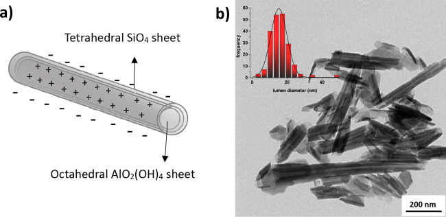
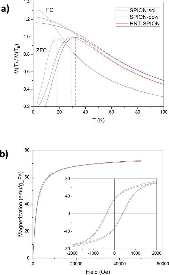
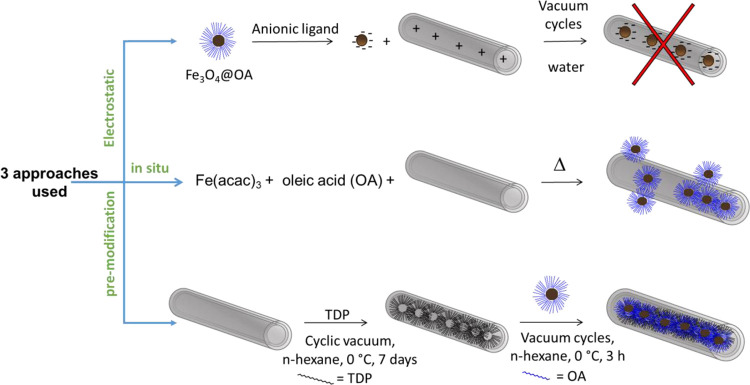
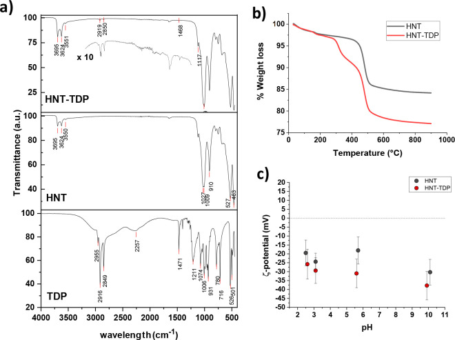
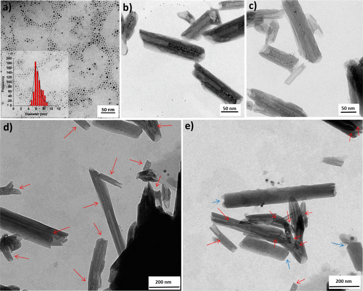
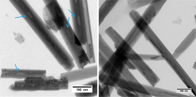
We present for the first time a method for the preparation of magnetic halloysite nanotubes (HNT) by loading of preformed superparamagnetic magnetite nanoparticles (SPION) of diameter size ∼6 nm with a hydrodynamic diameter of ∼10 nm into HNT. We found that the most effective route to reach this goal relies on the modification of the inner lumen of HNT by tetradecylphosphonic acid (TDP) to give HNT-TDP, followed by the loading with preformed oleic acid (OA)-stabilized SPION. Transmission electron microscopy evidenced the presence of highly crystalline magnetic nanoparticles only in the lumen, partially ordered in chainlike structures. Conversely, attempts to obtain the same result by exploiting either the positive charge of the HNT inner lumen employing SPIONs covered with negatively charged capping agents or the synthesis of SPION by thermal decomposition were not effective. HNT-TDP were characterized by infrared spectroscopy (ATR-FTIR), thermogravimetric analysis (TGA), and ζ-potential, and all of the techniques confirmed the presence of TDP onto the HNT. Moreover, the inner localization of TDP was ascertained by the use of Nile Red, a molecule whose luminescence is very sensitive to the polarity of the environment. The free SPION@OA (as a colloidal suspension and as a powder) and SPION-in-HNT powder were magnetically characterized by measuring the ZFC-FC magnetization curves as well as the hysteresis cycles at 300 and 2.5 K, confirming that the super-paramagnetic behavior and the main magnetic properties of the free SPION were preserved once embedded in SPION-in-HNT.
我们首次提出了一种通过将直径约为 6nm、水动力学直径约为 10nm 的超顺磁磁铁矿纳米粒子(SPION)负载到中空纳米管(HNT)内来制备磁性埃洛石纳米管(HNT)的方法。我们发现,实现这一目标的最有效途径是通过十四烷基膦酸(TDP)对 HNT 的内腔进行修饰,得到 HNT-TDP,然后负载预先形成的油酸(OA)稳定的 SPION。透射电子显微镜证明了只有在内腔中存在高度结晶的磁性纳米粒子,它们部分有序地排列成链状结构。相反,试图利用 HNT 内腔的正电荷,采用带有带负电荷的封端剂的 SPION,或者通过热分解合成 SPION 来获得相同的结果都没有效果。通过衰减全反射傅里叶变换红外光谱(ATR-FTIR)、热重分析(TGA)和 ζ-电位对 HNT-TDP 进行了表征,所有技术都证实了 TDP 存在于 HNT 上。此外,通过使用尼罗红(一种其发光对环境极性非常敏感的分子)证实了 TDP 的内腔定位。自由 SPION@OA(作为胶体悬浮液和粉末)和 SPION-in-HNT 粉末的磁性通过测量 ZFC-FC 磁化曲线以及在 300 和 2.5K 下的磁滞回线来进行表征,证实了自由 SPION 的超顺磁行为和主要磁性在嵌入 SPION-in-HNT 后得以保留。