National Resource Center for Non-human Primates, Kunming Primate Research Center, and National Research Facility for Phenotypic & Genetic Analysis of Model Animals (Primate Facility), Kunming Institute of Zoology, Chinese Academy of Sciences, Kunming, Yunnan, China.
University of Chinese Academy of Sciences, Beijing, China.
Cereb Cortex. 2021 Jan 1;31(1):341-355. doi: 10.1093/cercor/bhaa229.
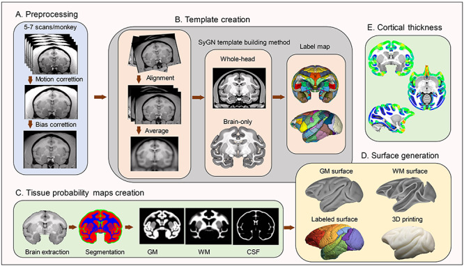
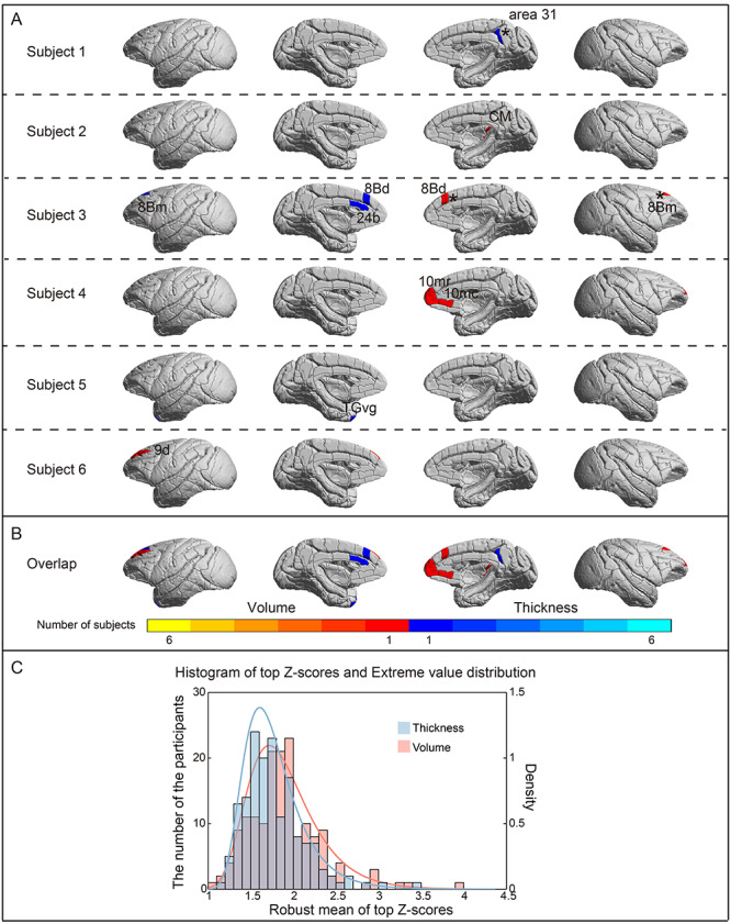
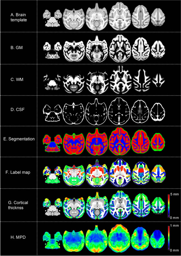
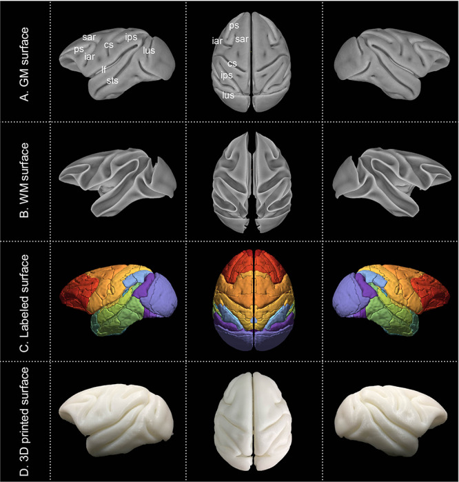
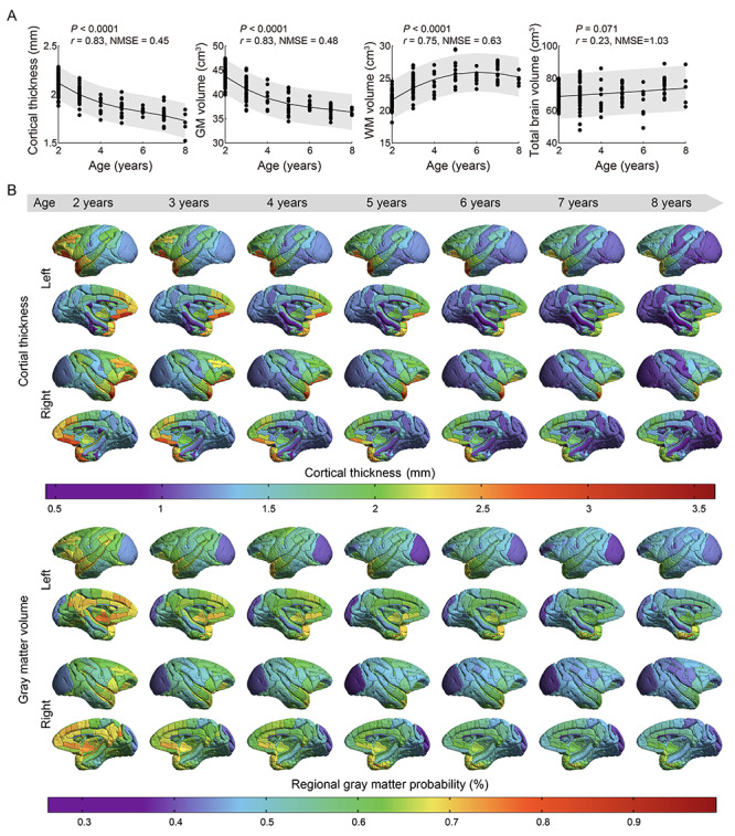
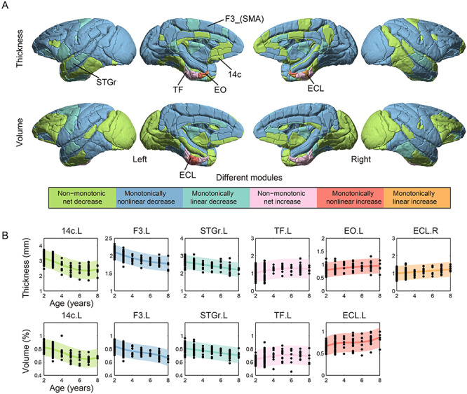
The developmental trajectory of the primate brain varies substantially with aging across subjects. However, this ubiquitous variability between individuals in brain structure is difficult to quantify and has thus essentially been ignored. Based on a large-scale structural magnetic resonance imaging dataset acquired from 162 cynomolgus macaques, we create a species-specific 3D template atlas of the macaque brain, and deploy normative modeling to characterize individual variations of cortical thickness (CT) and regional gray matter volume (GMV). We observed an overall decrease in total GMV and mean CT, and an increase in white matter volume from juvenile to early adult. Specifically, CT and regional GMV were greater in prefrontal and temporal cortices relative to early unimodal areas. Age-dependent trajectories of thickness and volume for each cortical region revealed an increase in the medial temporal lobe, and decreases in all other regions. A low percentage of highly individualized deviations of CT and GMV were identified (0.0021%, 0.0043%, respectively, P < 0.05, false discovery rate [FDR]-corrected). Our approach provides a natural framework to parse individual neuroanatomical differences for use as a reference standard in macaque brain research, potentially enabling inferences regarding the degree to which behavioral or symptomatic variables map onto brain structure in future disease studies.
灵长类动物大脑的发育轨迹在不同个体之间随年龄的变化而有很大差异。然而,这种个体间大脑结构的普遍可变性很难被量化,因此基本上被忽略了。基于从 162 只食蟹猴获得的大规模结构磁共振成像数据集,我们创建了一个特定于物种的猕猴大脑 3D 模板图谱,并部署了规范建模来描述皮质厚度 (CT) 和区域灰质体积 (GMV) 的个体变化。我们观察到从幼年到成年早期,总 GMV 和平均 CT 总体下降,而白质体积增加。具体而言,相对于早期单模态区域,前额叶和颞叶的 CT 和区域 GMV 更大。每个皮质区域的厚度和体积的年龄相关轨迹显示出内侧颞叶的增加,以及所有其他区域的减少。发现 CT 和 GMV 的高度个体化偏差的比例较低(分别为 0.0021%和 0.0043%,P<0.05,经 FDR 校正)。我们的方法为解析个体神经解剖差异提供了一个自然框架,可作为猕猴大脑研究的参考标准,可能使我们能够推断在未来的疾病研究中,行为或症状变量与大脑结构的对应程度。