Nollet Edgar E, Manders Emmy M, Goebel Max, Jansen Valentijn, Brockmann Cord, Osinga Jorrit, van der Velden Jolanda, Helmes Michiel, Kuster Diederik W D
Department of Physiology, Amsterdam UMC, Vrije Universiteit Amsterdam, Amsterdam Cardiovascular Sciences, Amsterdam, Netherlands.
CytoCypher BV, Wageningen, Netherlands.
Front Physiol. 2020 Jul 21;11:815. doi: 10.3389/fphys.2020.00815. eCollection 2020.
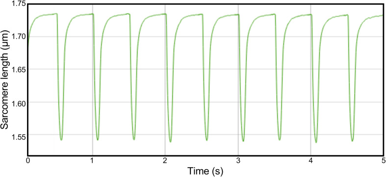
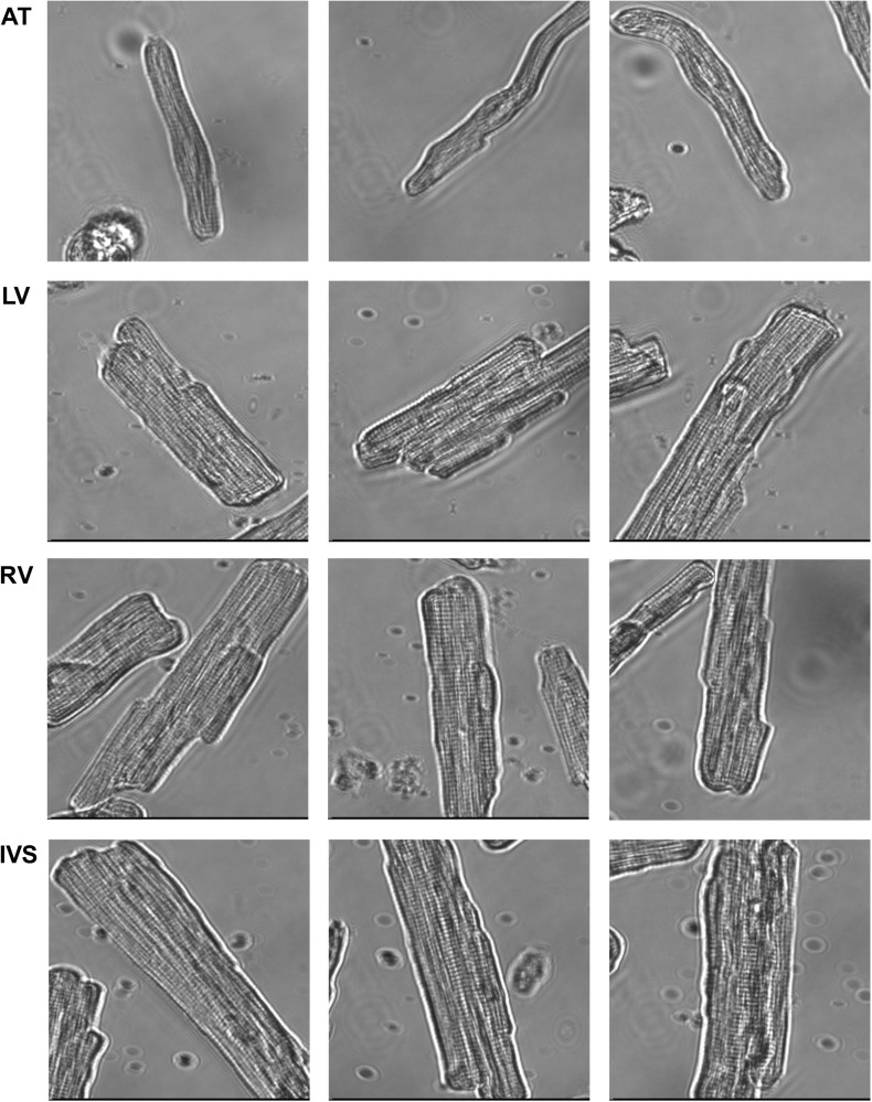
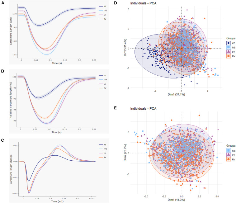
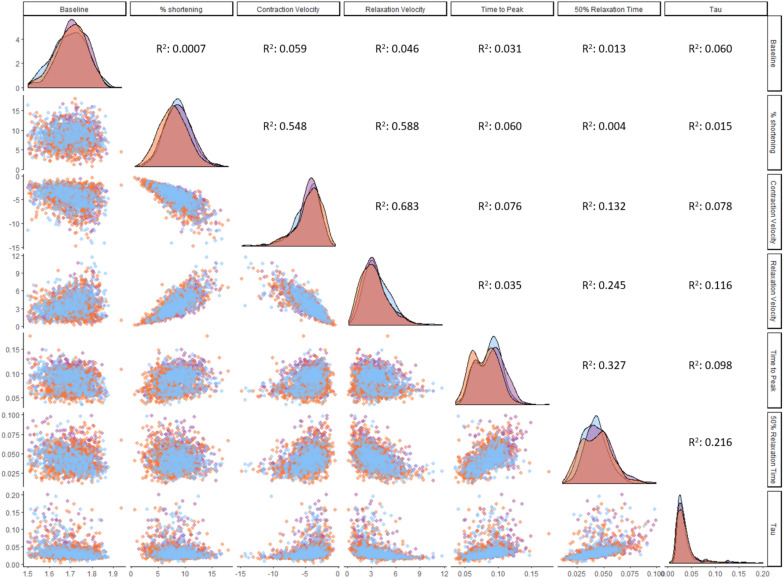
The chambers of the heart fulfill different hemodynamic functions, which are reflected in their structural and contractile properties. While the atria are highly elastic to allow filling from the venous system, the ventricles need to be able to produce sufficiently high pressures to eject blood into the circulation. The right ventricle (RV) pumps into the low pressure pulmonary circulation, while the left ventricle (LV) needs to overcome the high pressure of the systemic circulation. It is incompletely understood whether these differences can be explained by the contractile differences at the level of the individual cardiomyocytes of the chambers. We addressed this by isolating cardiomyocytes from atria, RV, LV, and interventricular septum (IVS) of five healthy wild-type rats. Using a high-throughput contractility set-up, we measured contractile function of 2,043 cells after overnight culture. Compared to ventricular cardiomyocytes, atrial cells showed a twofold lower contraction amplitude and 1.4- to 1.7-fold slower kinetics of contraction and relaxation. The interventricular differences in contractile function were much smaller; RV cells displayed 12-13% less fractional shortening and 5-9% slower contraction and 3-15% slower relaxation kinetics relative to their LV and IVS counterparts. Aided by a large dataset, we established relationships between contractile parameters and found contraction velocity, fractional shortening and relaxation velocity to be highly correlated. In conclusion, our findings are in line with contractile differences observed at the atrioventricular level, but can only partly explain the interventricular differences that exist at the organ level.
心脏的腔室具有不同的血液动力学功能,这在其结构和收缩特性中得以体现。心房具有高度弹性,以便从静脉系统接受血液充盈,而心室则需要能够产生足够高的压力,将血液泵入循环系统。右心室将血液泵入低压的肺循环,而左心室则需要克服体循环的高压。目前尚不完全清楚这些差异是否可以通过腔室中单个心肌细胞水平的收缩差异来解释。我们通过从五只健康野生型大鼠的心房、右心室、左心室和室间隔分离心肌细胞来解决这个问题。使用高通量收缩性检测装置,我们在过夜培养后测量了2043个细胞的收缩功能。与心室心肌细胞相比,心房细胞的收缩幅度低两倍,收缩和舒张动力学慢1.4至1.7倍。心室之间的收缩功能差异要小得多;相对于左心室和室间隔的对应细胞,右心室细胞的缩短分数少12 - 13%,收缩慢5 - 9%,舒张动力学慢3 - 15%。借助大量数据集,我们建立了收缩参数之间的关系,发现收缩速度、缩短分数和舒张速度高度相关。总之,我们的研究结果与在房室水平观察到的收缩差异一致,但只能部分解释器官水平存在的心室间差异。