Strome Arianna, Kossatz Susanne, Zanoni Daniella Karassawa, Rajadhyaksha Milind, Patel Snehal, Reiner Thomas
Department of Radiology, Memorial Sloan Kettering Cancer Center, New York, NY, USA.
Department of Surgery, Memorial Sloan Kettering Cancer Center, New York, NY, USA.
Mol Imaging. 2018 Jan-Dec;17:1536012118808644. doi: 10.1177/1536012118808644.
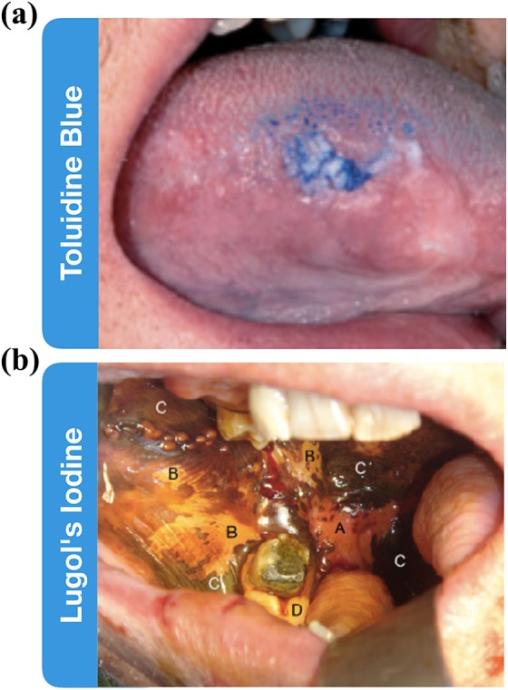
Oral cancer is one of the most common cancers globally. Survival rates for patients are directly correlated with stage of diagnosis; despite this knowledge, 60% of individuals are presenting with late-stage disease. Currently, the initial evaluation of a questionable lesion is performed by a conventional visual examination with white light. If a lesion is deemed suspicious, a biopsy is taken for diagnosis. However, not all lesions present suspicious under visual white light examination, and there is limited specificity in differentiating between benign and malignant transformations. Several vital dyes, light-based detection systems, and cytology evaluation methods have been formulated to aid in the visualization process, but their lack of specific biomarkers resulted in high false-positive rates and thus limits their reliability as screening and guidance tools. In this review, we will analyze the current methodologies and demonstrate the need for specific intraoral imaging agents to aid in screening and diagnosis to identify patients earlier. Several novel molecular imaging agents will be presented as, by result of their molecular targeting, they aim to have high specificity for tumor pathways and can support in identifying dysplastic/cancerous lesions and guiding visualization of biopsy sites. Imaging agents that are easy to use, inexpensive, noninvasive, and specific can be utilized to increase the number of patients who are screened and monitored in a variety of different environments, with the ultimate goal of increasing early detection.
口腔癌是全球最常见的癌症之一。患者的生存率与诊断阶段直接相关;尽管有这一认识,但60%的患者就诊时已处于疾病晚期。目前,对可疑病变的初步评估是通过白光下的传统视觉检查进行的。如果病变被认为可疑,则进行活检以确诊。然而,并非所有病变在视觉白光检查下都表现出可疑特征,并且在区分良性和恶性病变方面特异性有限。已经开发了几种活性染料、基于光的检测系统和细胞学评估方法来辅助可视化过程,但它们缺乏特异性生物标志物导致假阳性率高,因此限制了它们作为筛查和指导工具的可靠性。在本综述中,我们将分析当前的方法,并证明需要特定的口腔内成像剂来辅助筛查和诊断,以便更早地识别患者。将介绍几种新型分子成像剂,由于它们的分子靶向作用,旨在对肿瘤通路具有高特异性,并可支持识别发育异常/癌性病变以及指导活检部位的可视化。易于使用、价格低廉、无创且具有特异性的成像剂可用于增加在各种不同环境中接受筛查和监测的患者数量,最终目标是提高早期检测率。