Department of Pathology, University of California San Francisco, San Francisco, CA, USA.
Pathology Service 113B, San Francisco VA Health Care System, San Francisco, CA, USA.
Nature. 2020 Dec;588(7838):459-465. doi: 10.1038/s41586-020-2709-7. Epub 2020 Aug 31.
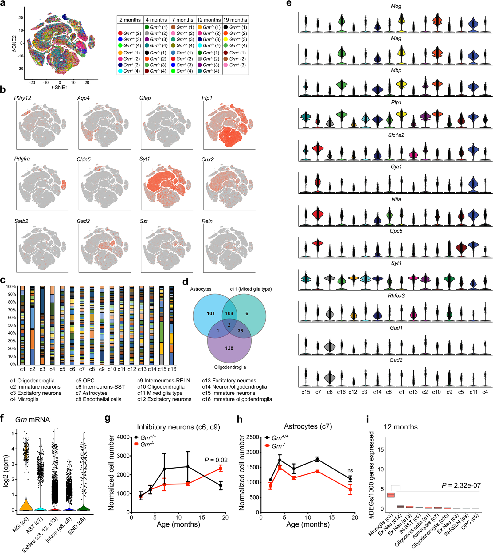
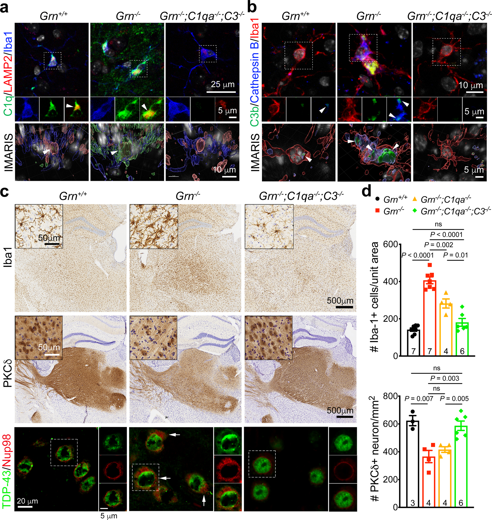
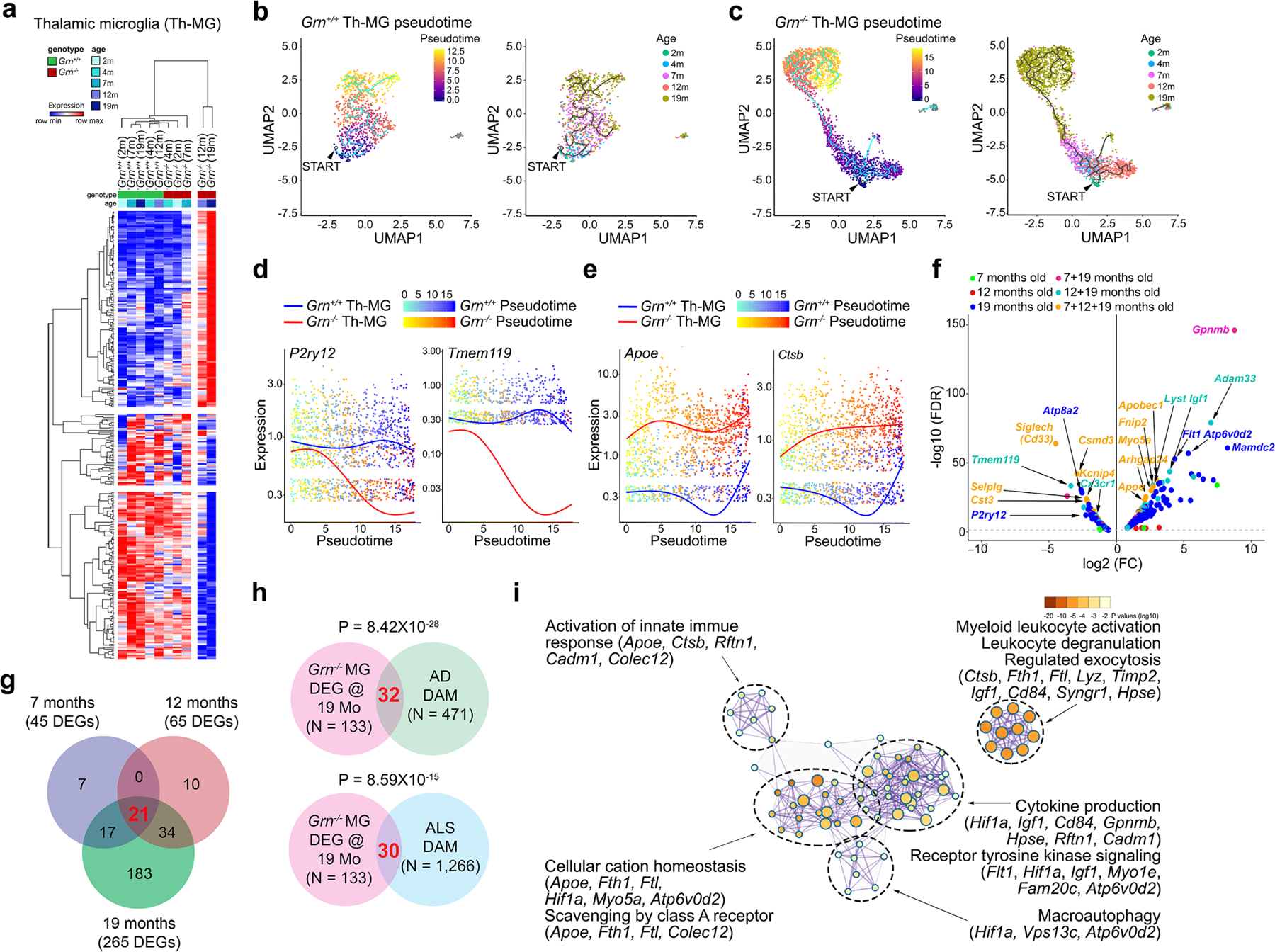
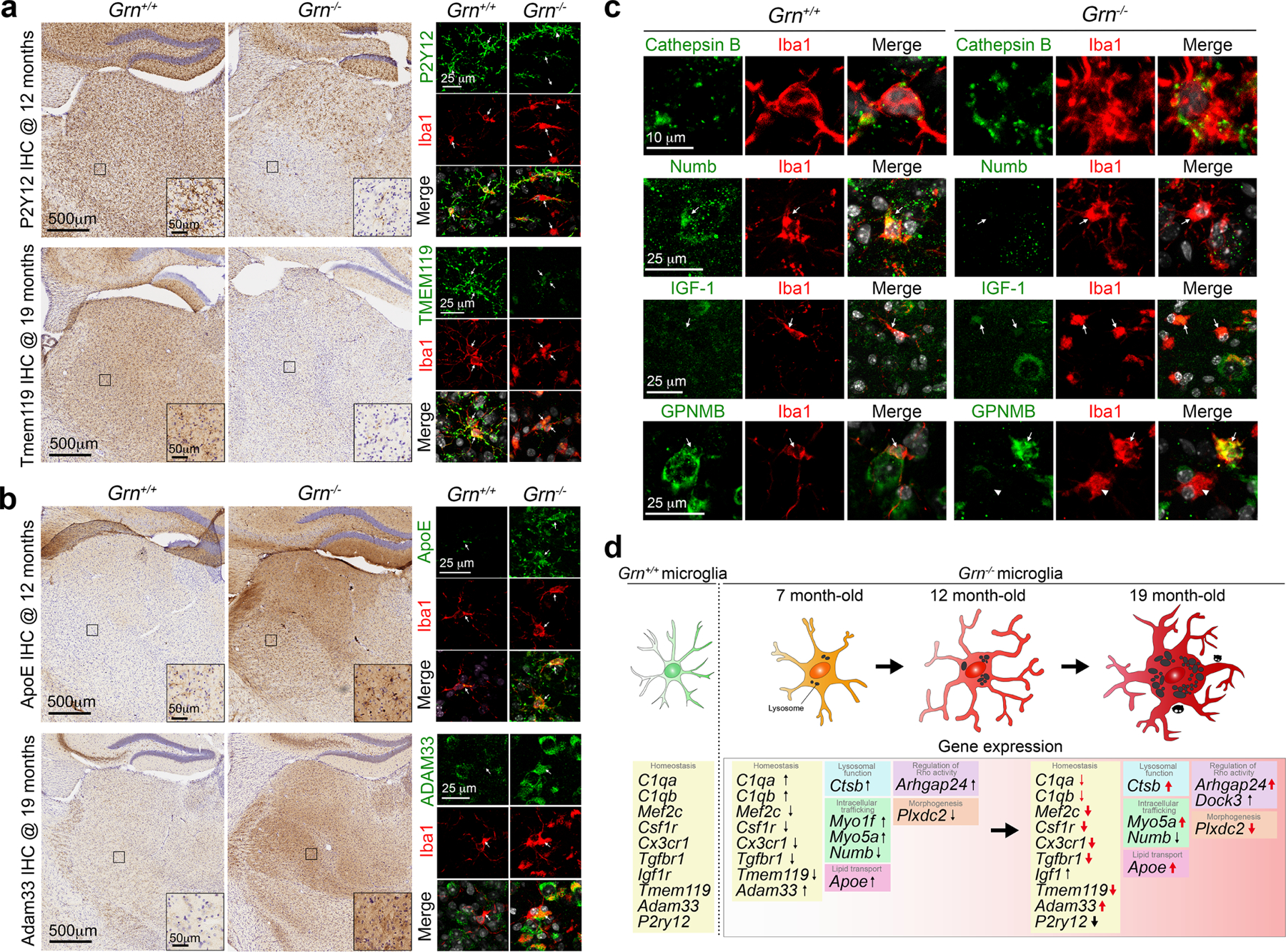
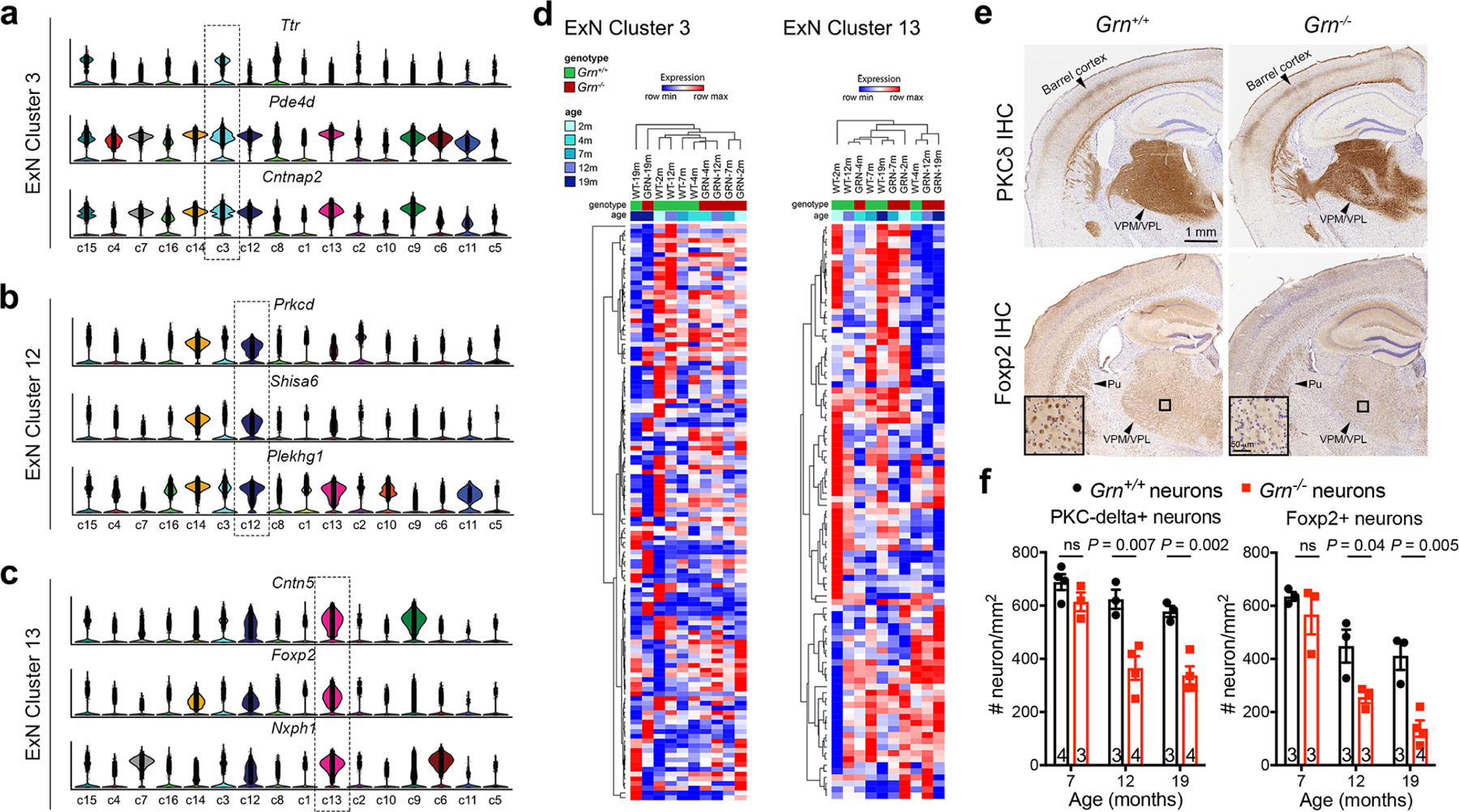
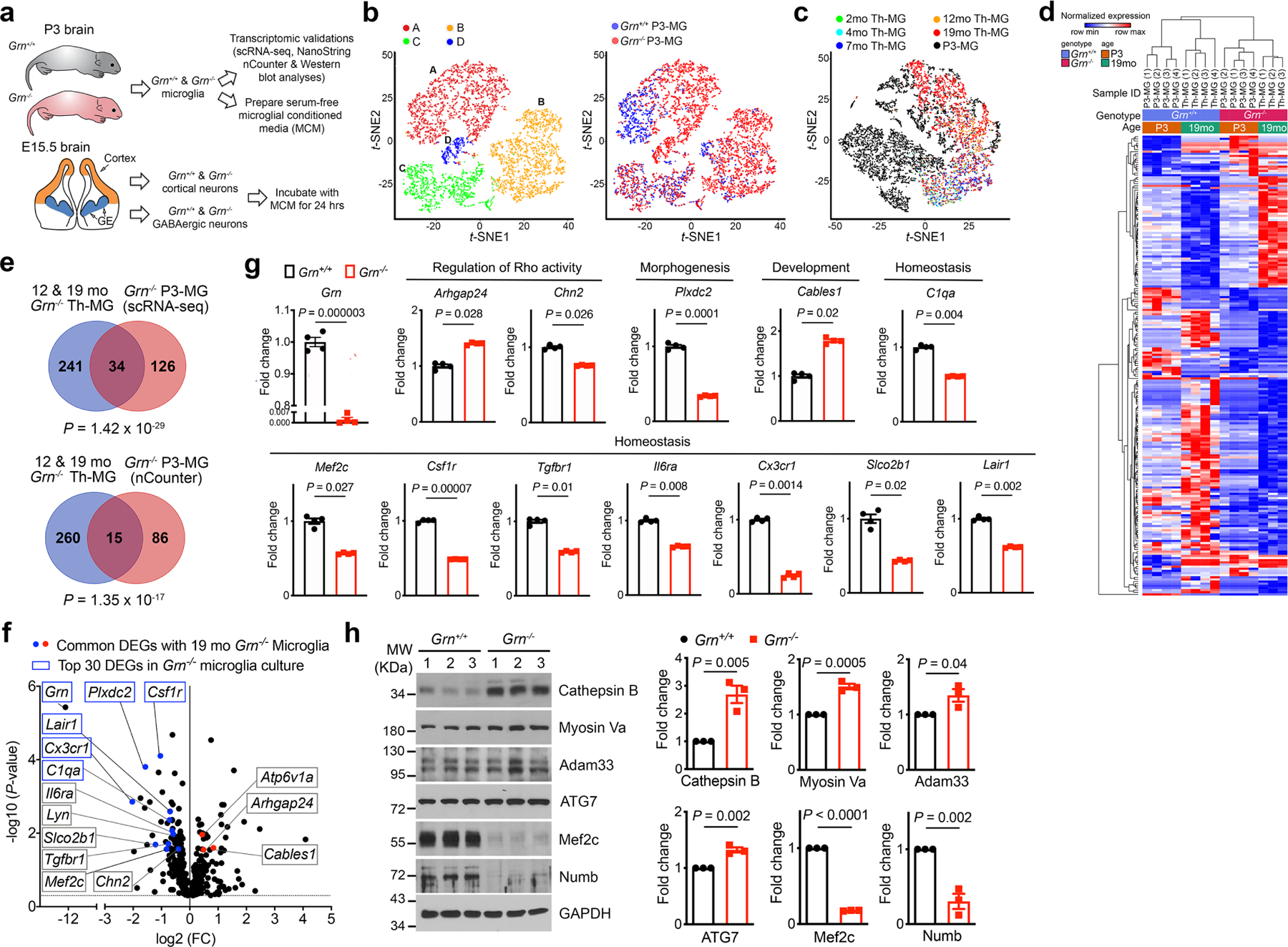
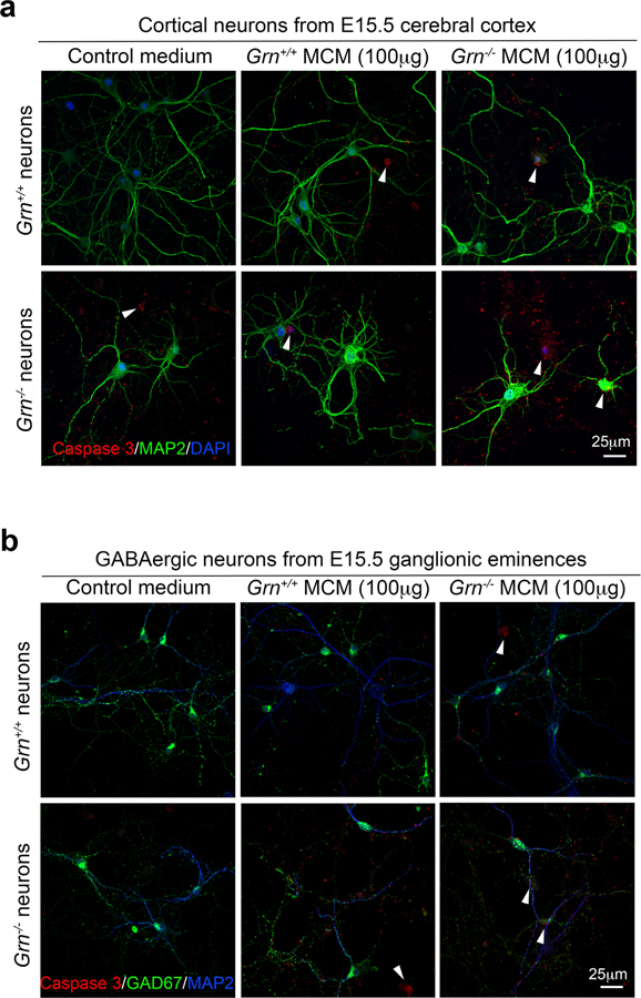
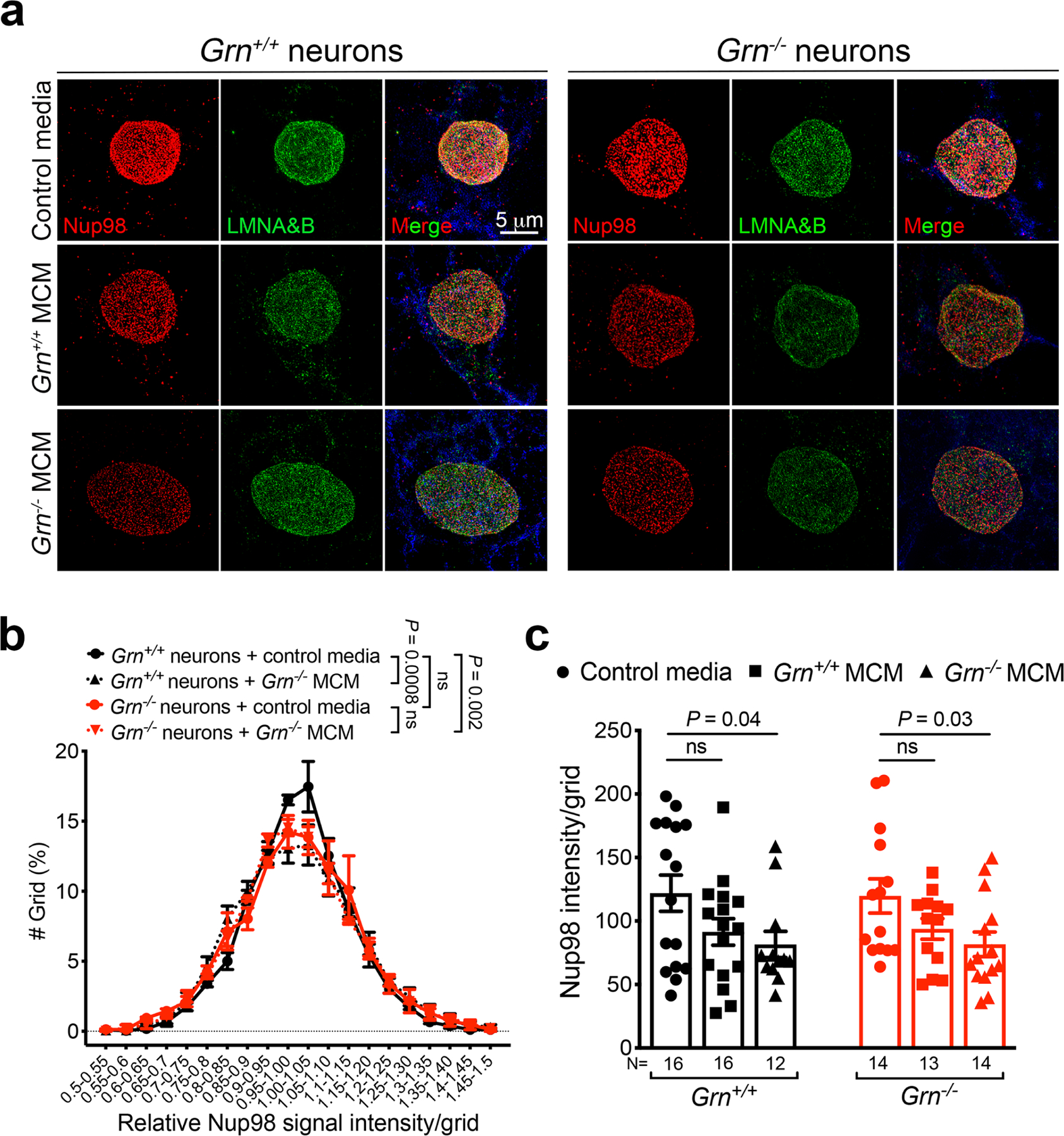
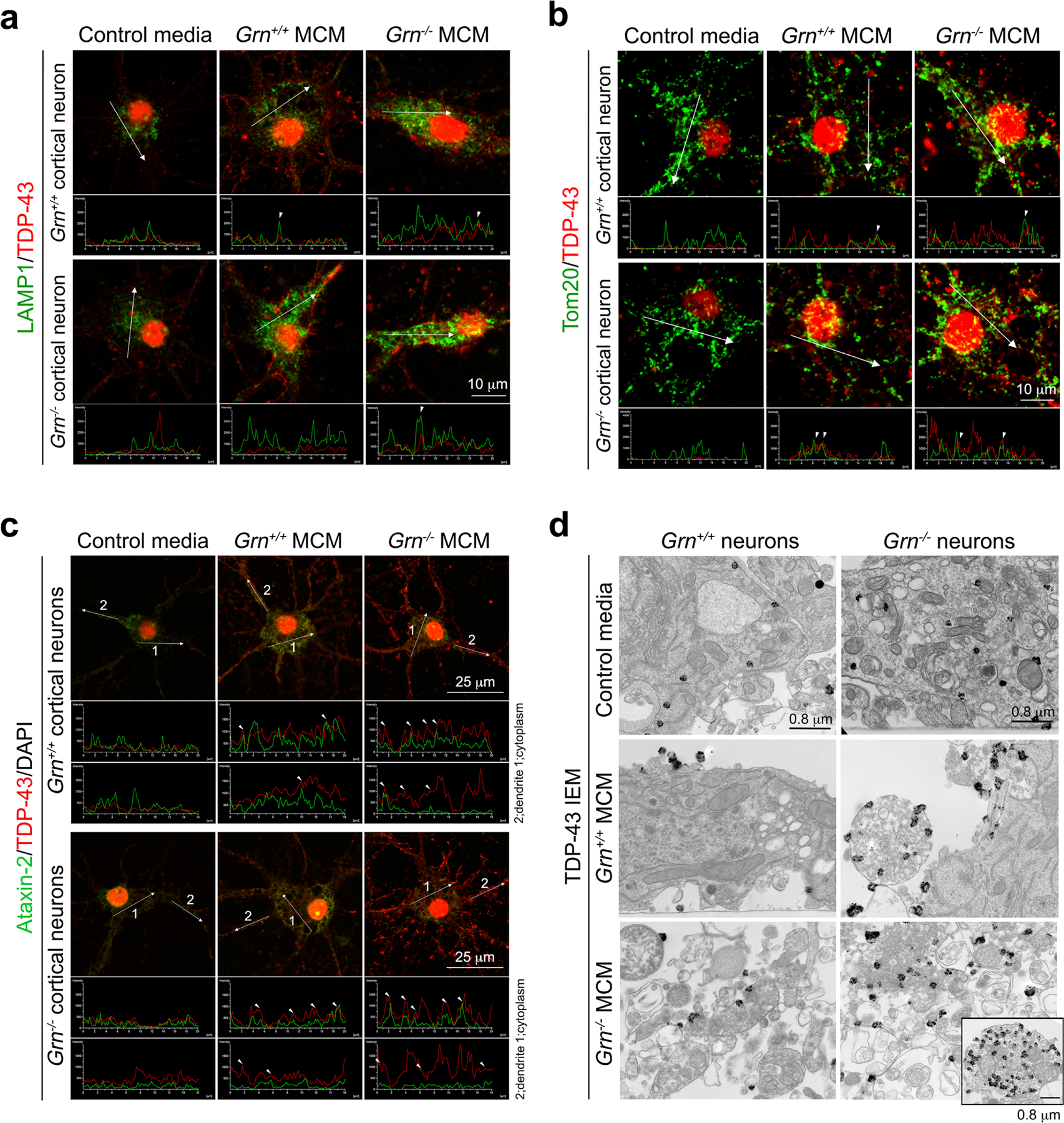
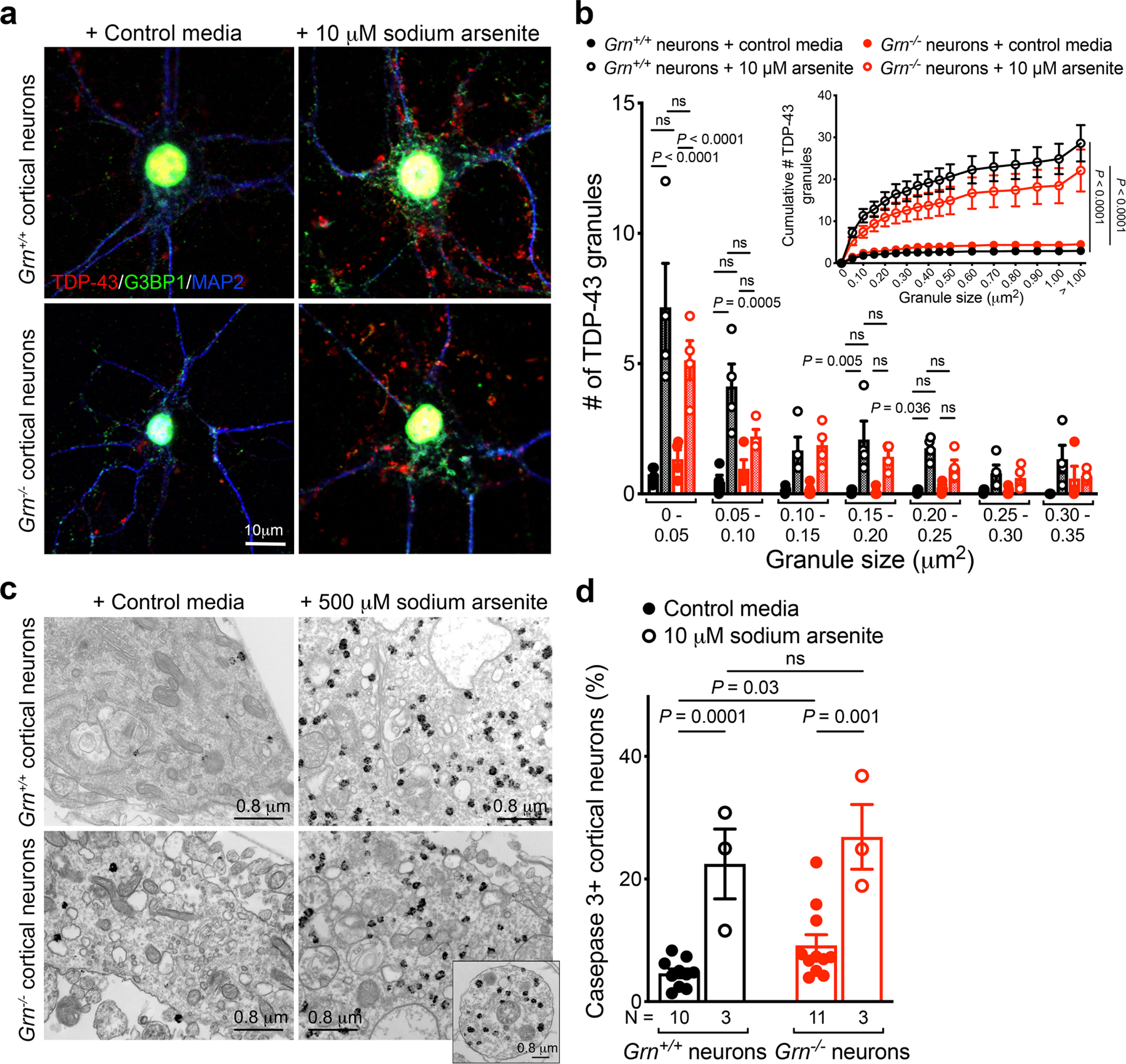
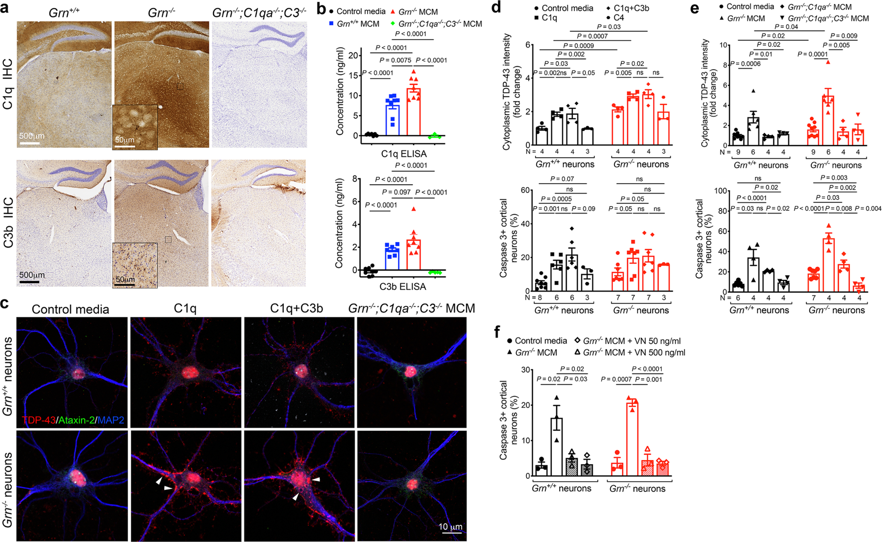
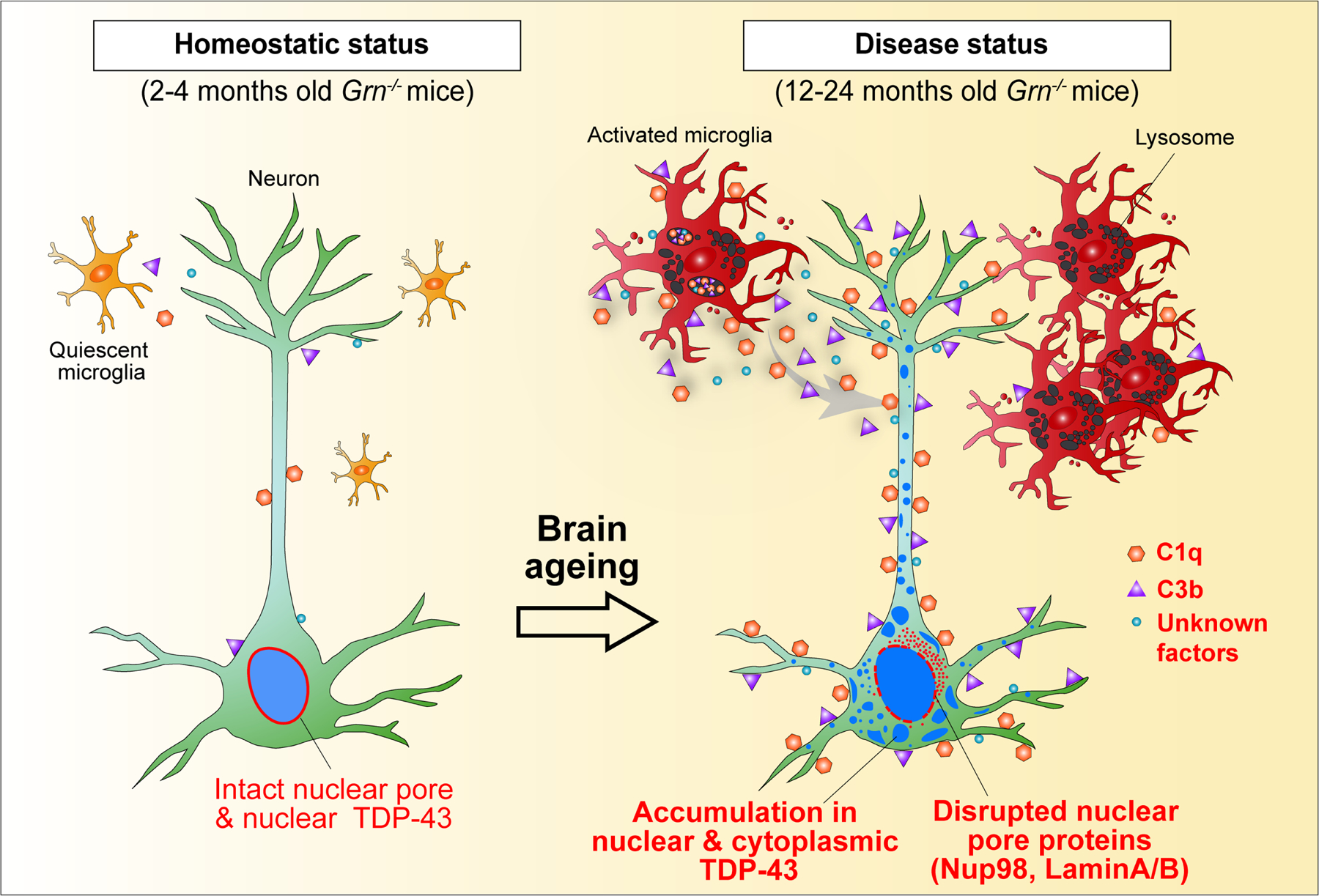
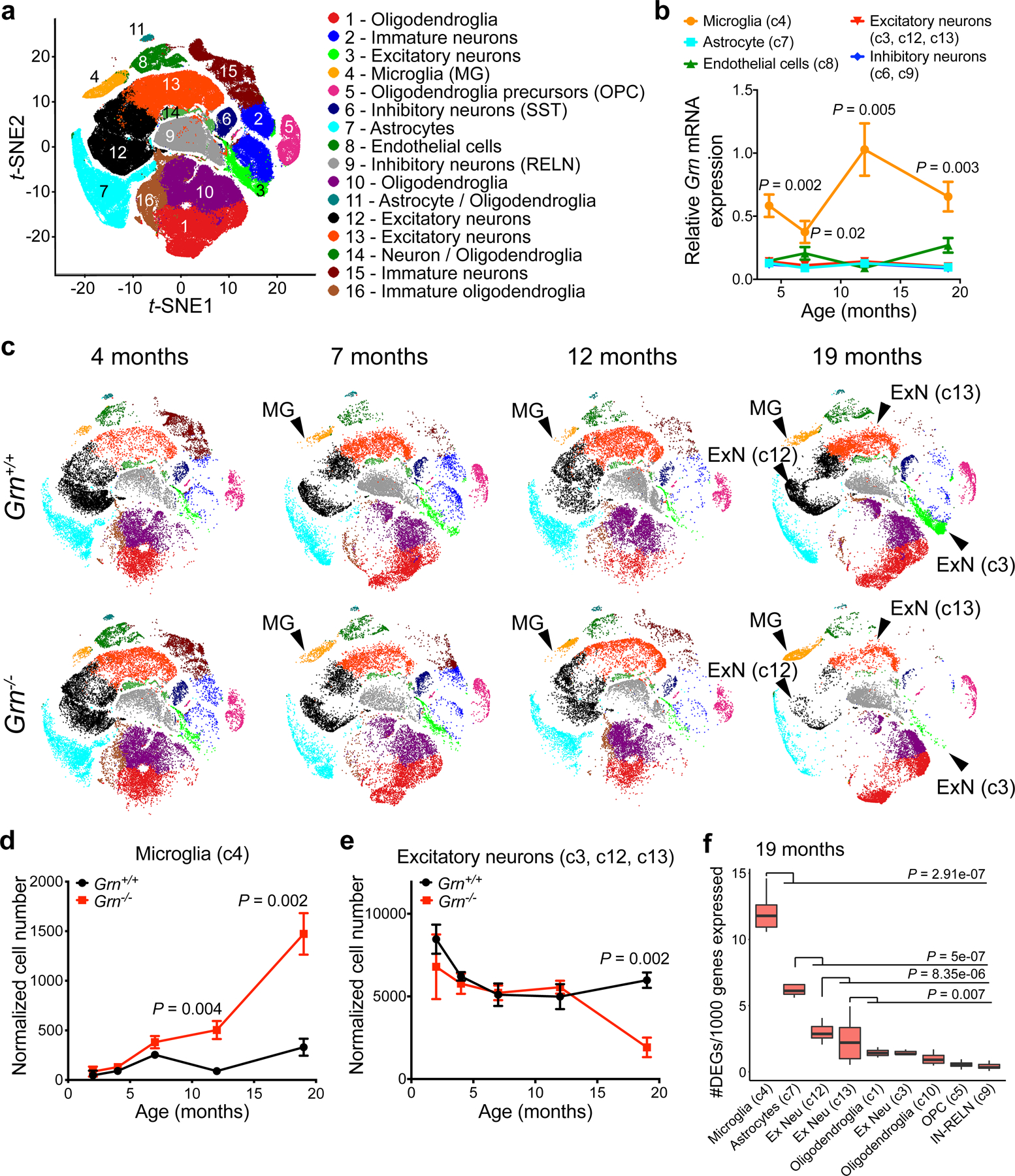
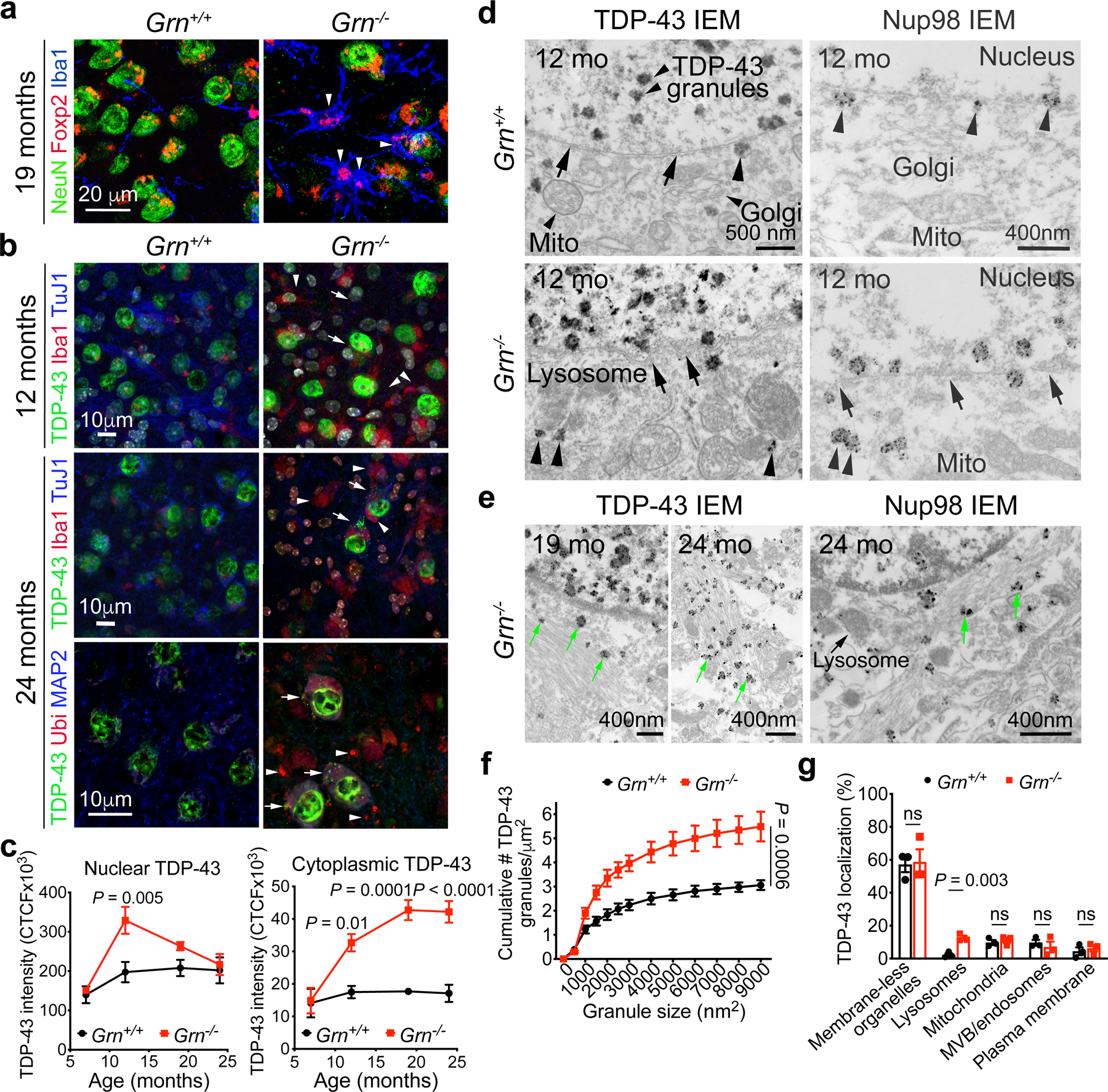
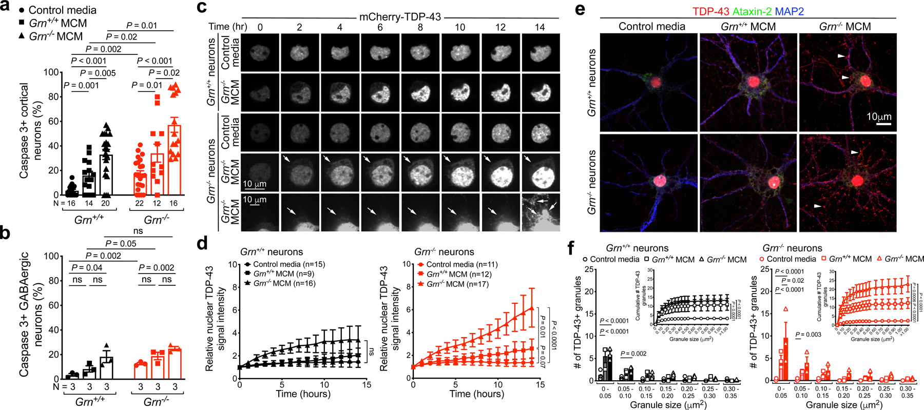
Aberrant aggregation of the RNA-binding protein TDP-43 in neurons is a hallmark of frontotemporal lobar degeneration caused by haploinsufficiency in the gene encoding progranulin. However, the mechanism leading to TDP-43 proteinopathy remains unclear. Here we use single-nucleus RNA sequencing to show that progranulin deficiency promotes microglial transition from a homeostatic to a disease-specific state that causes endolysosomal dysfunction and neurodegeneration in mice. These defects persist even when Grn microglia are cultured ex vivo. In addition, single-nucleus RNA sequencing reveals selective loss of excitatory neurons at disease end-stage, which is characterized by prominent nuclear and cytoplasmic TDP-43 granules and nuclear pore defects. Remarkably, conditioned media from Grn microglia are sufficient to promote TDP-43 granule formation, nuclear pore defects and cell death in excitatory neurons via the complement activation pathway. Consistent with these results, deletion of the genes encoding C1qa and C3 mitigates microglial toxicity and rescues TDP-43 proteinopathy and neurodegeneration. These results uncover previously unappreciated contributions of chronic microglial toxicity to TDP-43 proteinopathy during neurodegeneration.
TDP-43 蛋白在神经元中的异常聚集是由编码颗粒蛋白前体的基因单倍体不足引起的额颞叶退行性变的一个标志。然而,导致 TDP-43 蛋白病的机制仍不清楚。在这里,我们使用单核 RNA 测序表明,颗粒蛋白缺乏会促进小胶质细胞从稳态向疾病特异性状态的转变,从而导致小鼠内溶酶体功能障碍和神经退行性变。即使在体外培养 Grn 小胶质细胞时,这些缺陷仍然存在。此外,单核 RNA 测序揭示了疾病终末期兴奋性神经元的选择性丧失,其特征是核内和细胞质 TDP-43 颗粒明显增多和核孔缺陷。值得注意的是,Grn 小胶质细胞的条件培养基通过补体激活途径足以促进兴奋性神经元中 TDP-43 颗粒的形成、核孔缺陷和细胞死亡。与这些结果一致的是,缺失编码 C1qa 和 C3 的基因减轻了小胶质细胞的毒性,并挽救了 TDP-43 蛋白病和神经退行性变。这些结果揭示了慢性小胶质细胞毒性对神经退行性变过程中 TDP-43 蛋白病的先前未被认识到的贡献。