• 文献检索
  • 文档翻译
  • 深度研究
  • 学术资讯
  • Suppr Zotero 插件Zotero 插件
  • 邀请有礼
  • 套餐&价格
  • 历史记录
应用&插件
Suppr Zotero 插件Zotero 插件浏览器插件Mac 客户端Windows 客户端微信小程序
定价
高级版会员购买积分包购买API积分包
服务
文献检索文档翻译深度研究API 文档MCP 服务
关于我们
关于 Suppr公司介绍联系我们用户协议隐私条款
关注我们

Suppr 超能文献

核心技术专利:CN118964589B侵权必究
粤ICP备2023148730 号-1Suppr @ 2026

文献检索

告别复杂PubMed语法,用中文像聊天一样搜索,搜遍4000万医学文献。AI智能推荐,让科研检索更轻松。

立即免费搜索

文件翻译

保留排版,准确专业,支持PDF/Word/PPT等文件格式,支持 12+语言互译。

免费翻译文档

深度研究

AI帮你快速写综述,25分钟生成高质量综述,智能提取关键信息,辅助科研写作。

立即免费体验

肌肉骨骼超声检查中的伪像:从物理到临床

Artifacts in Musculoskeletal Ultrasonography: From Physics to Clinics.

作者信息

Wu Wei-Ting, Chang Ke-Vin, Hsu Yu-Chun, Hsu Po-Cheng, Ricci Vincenzo, Özçakar Levent

机构信息

Department of Physical Medicine and Rehabilitation, National Taiwan University Hospital, Bei-Hu Branch, Taipei 10845, Taiwan.

Department of Physical Medicine and Rehabilitation, National Taiwan University College of Medicine, Taipei 10048, Taiwan.

出版信息

Diagnostics (Basel). 2020 Aug 27;10(9):645. doi: 10.3390/diagnostics10090645.

DOI:10.3390/diagnostics10090645
PMID:32867385
原文链接:https://pmc.ncbi.nlm.nih.gov/articles/PMC7555047/
Abstract

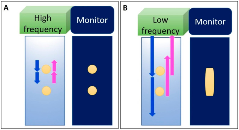
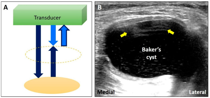
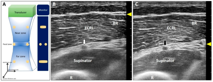
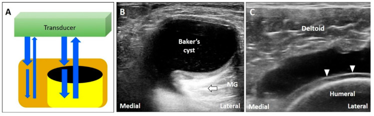
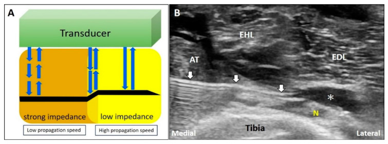
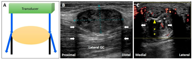
Ultrasound appears to be the most useful imaging tool in the diagnosis and guided treatment of musculoskeletal disorders. However, ultrasonography has been criticized for being user dependent. Therefore, medical professionals should be familiar with the basic principles of ultrasound imaging (e.g., physics and technical skills) to diminish artifacts and avoid misinterpretation. In this review, we focused on the physics of common artifacts, their clinical significance, and the ways to tackle them in daily practice during musculoskeletal imaging. In particular, artifacts pertaining to the focal zone, beam attenuation, path and side lobe of the beam, speed of the sound, and range ambiguity were described.

摘要

超声似乎是肌肉骨骼疾病诊断和引导治疗中最有用的成像工具。然而,超声检查因依赖操作者而受到批评。因此,医学专业人员应熟悉超声成像的基本原理(如物理和技术技能),以减少伪像并避免误判。在本综述中,我们重点关注常见伪像的物理原理、它们的临床意义以及在肌肉骨骼成像日常实践中处理这些伪像的方法。特别描述了与聚焦区、波束衰减、波束路径和旁瓣、声速以及距离模糊有关的伪像。

https://cdn.ncbi.nlm.nih.gov/pmc/blobs/6618/7555047/586ad871178d/diagnostics-10-00645-g014.jpg
https://cdn.ncbi.nlm.nih.gov/pmc/blobs/6618/7555047/14fecc7ab0dd/diagnostics-10-00645-g001.jpg
https://cdn.ncbi.nlm.nih.gov/pmc/blobs/6618/7555047/59af7c8fe020/diagnostics-10-00645-g002.jpg
https://cdn.ncbi.nlm.nih.gov/pmc/blobs/6618/7555047/9e3bc631bcda/diagnostics-10-00645-g003.jpg
https://cdn.ncbi.nlm.nih.gov/pmc/blobs/6618/7555047/845efafb343f/diagnostics-10-00645-g004.jpg
https://cdn.ncbi.nlm.nih.gov/pmc/blobs/6618/7555047/fb026df392c4/diagnostics-10-00645-g005.jpg
https://cdn.ncbi.nlm.nih.gov/pmc/blobs/6618/7555047/97c95c088032/diagnostics-10-00645-g006.jpg
https://cdn.ncbi.nlm.nih.gov/pmc/blobs/6618/7555047/ccfd964f3b7b/diagnostics-10-00645-g007.jpg
https://cdn.ncbi.nlm.nih.gov/pmc/blobs/6618/7555047/849eb42aa246/diagnostics-10-00645-g008.jpg
https://cdn.ncbi.nlm.nih.gov/pmc/blobs/6618/7555047/e09a3f117021/diagnostics-10-00645-g009.jpg
https://cdn.ncbi.nlm.nih.gov/pmc/blobs/6618/7555047/281f754d0e76/diagnostics-10-00645-g010.jpg
https://cdn.ncbi.nlm.nih.gov/pmc/blobs/6618/7555047/e81e55bdef12/diagnostics-10-00645-g011.jpg
https://cdn.ncbi.nlm.nih.gov/pmc/blobs/6618/7555047/b3a4558413be/diagnostics-10-00645-g012.jpg
https://cdn.ncbi.nlm.nih.gov/pmc/blobs/6618/7555047/480a3cd2a394/diagnostics-10-00645-g013.jpg
https://cdn.ncbi.nlm.nih.gov/pmc/blobs/6618/7555047/586ad871178d/diagnostics-10-00645-g014.jpg

相似文献

1
Artifacts in Musculoskeletal Ultrasonography: From Physics to Clinics.肌肉骨骼超声检查中的伪像:从物理到临床
Diagnostics (Basel). 2020 Aug 27;10(9):645. doi: 10.3390/diagnostics10090645.
2
Clinical Significance of US Artifacts.超声伪像的临床意义。
Radiographics. 2017 Sep-Oct;37(5):1408-1423. doi: 10.1148/rg.2017160175. Epub 2017 Aug 4.
3
Artifacts in ultrasound imaging.超声成像中的伪像。
J Ultrasound Med. 1986 Apr;5(4):227-37. doi: 10.7863/jum.1986.5.4.227.
4
[Ultrasonic imaging artifacts. Physical bases].[超声成像伪像。物理基础]
Radiologe. 1993 Jan;33(1):1-10.
5
Review of artifacts associated with transrectal ultrasound: understanding, recognition, and prevention of misinterpretation.经直肠超声相关伪像综述:对误判的理解、识别及预防
J Clin Ultrasound. 1995 Oct;23(8):483-94. doi: 10.1002/jcu.1870230805.
6
Contrast-Enhanced Sonography of the Liver: How to Avoid Artifacts.肝脏超声造影:如何避免伪像
Diagnostics (Basel). 2024 Aug 20;14(16):1817. doi: 10.3390/diagnostics14161817.
7
[Basic physical principles of ultrasonography, anatomy of the musculoskeletal system and ecographic artifacts].[超声检查的基本物理原理、肌肉骨骼系统解剖及超声伪像]
Acta Ortop Mex. 2008 Nov-Dec;22(6):361-73.
8
Physics and principles of breast ultrasound.乳腺超声的物理学原理
Am Surg. 1996 Feb;62(2):103-7.
9
Artifacts and Artistic Facts: A Visual Simulation for Ultrasound Training.伪影与艺术事实:超声培训的可视化模拟。
Am J Phys Med Rehabil. 2019 Jun;98(6):521-525. doi: 10.1097/PHM.0000000000001134.
10
US artifacts.美国伪影。
Radiographics. 2009 Jul-Aug;29(4):1179-89. doi: 10.1148/rg.294085199.

引用本文的文献

1
The Role of Ultrasound in Postoperative Assessment of Biceps Tenodesis: A Case Report.超声在肱二头肌肌腱固定术后评估中的作用:一例报告
Cureus. 2025 Jun 15;17(6):e86099. doi: 10.7759/cureus.86099. eCollection 2025 Jun.
2
Ultrasound Findings of Emphysematous Pyelonephritis: A Case Series.气肿性肾盂肾炎的超声表现:病例系列
J Am Coll Emerg Physicians Open. 2025 Jun 11;6(4):100190. doi: 10.1016/j.acepjo.2025.100190. eCollection 2025 Aug.
3
Wearable Ultrasound-Imaging-Based Visual Feedback (UVF) Training for Ankle Rehabilitation of Chronic Stroke Survivors: A Proof-of-Concept Randomized Crossover Study.

本文引用的文献

1
Advanced Musculoskeletal Ultrasound Techniques: What Are the Applications?高级肌肉骨骼超声技术:有哪些应用?
AJR Am J Roentgenol. 2021 Feb;216(2):436-445. doi: 10.2214/AJR.20.22840. Epub 2020 Dec 2.
2
Ultrasonographic evaluation of the patellar tendon length and elasticity after open-wedge high tibial osteotomy: A comparison with radiological and clinical parameters.经开放楔形胫骨高位截骨术后髌腱长度和弹性的超声评估:与放射学和临床参数的比较。
Knee. 2020 Aug;27(4):1128-1134. doi: 10.1016/j.knee.2020.05.016. Epub 2020 Jun 25.
3
Imaging of gout: findings and pitfalls. A pictorial review.
基于可穿戴超声成像的视觉反馈(UVF)训练用于慢性中风幸存者的踝关节康复:一项概念验证随机交叉研究
Biosensors (Basel). 2025 Jun 6;15(6):365. doi: 10.3390/bios15060365.
4
The (ProteUS) Anisotropy Effect in Deep Fascia Ultrasonography: The Impact of Probe Angulation on Echogenicity and Thickness Assessments.深筋膜超声检查中的(ProteUS)各向异性效应:探头角度对回声性和厚度评估的影响。
Life (Basel). 2025 May 21;15(5):822. doi: 10.3390/life15050822.
5
Ultrasonographic Evaluation of Solid Organ Sizes in Children with Primary Malnutrition: A Preliminary Study.原发性营养不良患儿实体器官大小的超声评估:一项初步研究。
J Clin Med. 2024 Dec 31;14(1):169. doi: 10.3390/jcm14010169.
6
Diagnostic Musculoskeletal Ultrasound for Medial Collateral Ligament Injuries: Applications in Rehabilitation.诊断性肌肉骨骼超声在膝关节内侧副韧带损伤中的应用:康复中的应用
Int J Sports Phys Ther. 2024 Sep 1;19(9):1166-1171. doi: 10.26603/001c.122931. eCollection 2024.
7
Current status of dynamic musculoskeletal ultrasound for application to treatment of orthopedic diseases.动态肌肉骨骼超声在骨科疾病治疗中的应用现状
Am J Transl Res. 2024 Jun 15;16(6):2180-2189. doi: 10.62347/WHER3512. eCollection 2024.
8
Ultrasound imaging of the axilla.腋窝的超声成像。
Insights Imaging. 2023 May 11;14(1):78. doi: 10.1186/s13244-023-01430-9.
9
Ultrasound Imaging and Guidance for Cervical Myofascial Pain: A Narrative Review.超声成像在颈肌筋膜疼痛中的应用:叙述性综述。
Int J Environ Res Public Health. 2023 Feb 21;20(5):3838. doi: 10.3390/ijerph20053838.
10
Ultrasound imaging of bone fractures.骨折的超声成像
Insights Imaging. 2022 Dec 13;13(1):189. doi: 10.1186/s13244-022-01335-z.
痛风的影像学表现:征象与陷阱。影像学图片解读。
Acta Reumatol Port. 2020 Jan-Mar;45(1):20-25.
4
Post-operative MRI and US appearance of the Achilles tendons.跟腱术后的 MRI 和超声表现。
J Ultrasound. 2020 Sep;23(3):387-395. doi: 10.1007/s40477-020-00479-2. Epub 2020 Jun 1.
5
Ultrasound Findings in Less Frequent Causes of Carpal Tunnel Syndrome.腕管综合征少见病因的超声表现
J Ultrasound Med. 2020 Dec;39(12):2469-2482. doi: 10.1002/jum.15349. Epub 2020 May 27.
6
Ultrasound and bone: a pictorial review.超声与骨骼:影像学综述。
J Ultrasound. 2020 Sep;23(3):227-257. doi: 10.1007/s40477-020-00477-4. Epub 2020 May 17.
7
Ultrasound of the ulnar-palmar region of the wrist: normal anatomy and anatomic variations.腕关节尺侧-掌侧区域的超声:正常解剖结构与解剖变异。
J Ultrasound. 2020 Sep;23(3):365-378. doi: 10.1007/s40477-020-00468-5. Epub 2020 May 8.
8
Advanced Ankle and Foot Sonoanatomy: Imaging Beyond the Basics.高级踝关节和足部超声解剖学:超越基础的成像技术
Diagnostics (Basel). 2020 Mar 14;10(3):160. doi: 10.3390/diagnostics10030160.
9
Clinical Tests of the Shoulder: Accuracy and Extension Using Dynamic Ultrasound.肩部临床检查:动态超声的准确性和应用范围。
Am J Phys Med Rehabil. 2020 Feb;99(2):161-169. doi: 10.1097/PHM.0000000000001311.
10
Ulnar Wrist Pain Revisited: Ultrasound Diagnosis and Guided Injection for Triangular Fibrocartilage Complex Injuries.尺侧腕部疼痛再探讨:三角纤维软骨复合体损伤的超声诊断与引导下注射治疗
J Clin Med. 2019 Sep 25;8(10):1540. doi: 10.3390/jcm8101540.