Department of Pediatrics, Washington University School of Medicine, St. Louis, MO, USA.
Department of Neurology, Washington University School of Medicine, St. Louis, MO, USA.
Ann Neurol. 2020 Nov;88(5):995-1008. doi: 10.1002/ana.25891. Epub 2020 Sep 16.
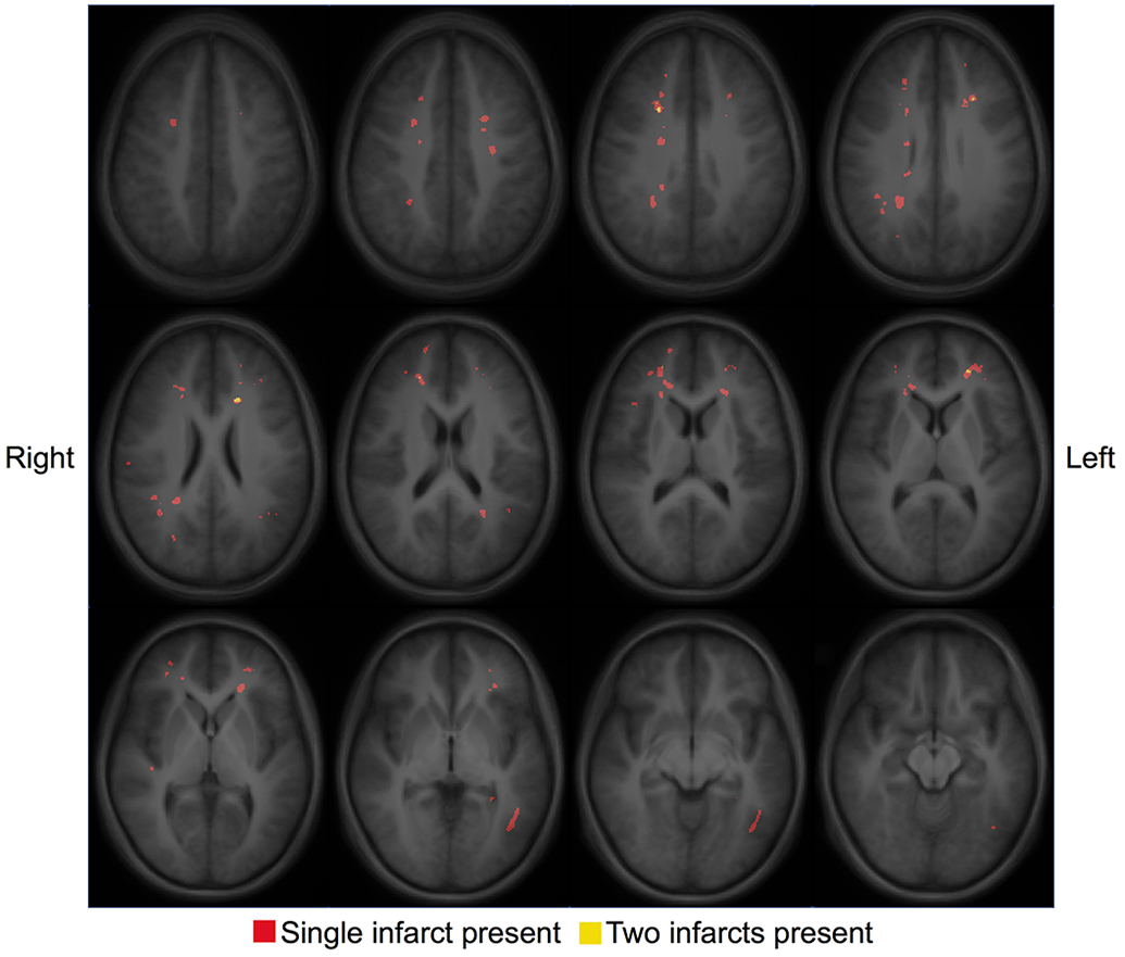
Children with sickle cell disease (SCD) experience cognitive deficits even when unaffected by stroke. Using functional connectivity magnetic resonance imaging (MRI) as a potential biomarker of cognitive function, we tested our hypothesis that children with SCD would have decreased functional connectivity, and that children experiencing the greatest metabolic stress, indicated by elevated oxygen extraction fraction, would have the lowest connectivity.
We prospectively obtained brain MRIs and cognitive testing in healthy controls and children with SCD.
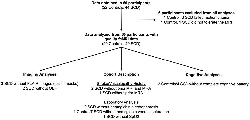
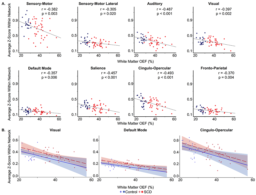
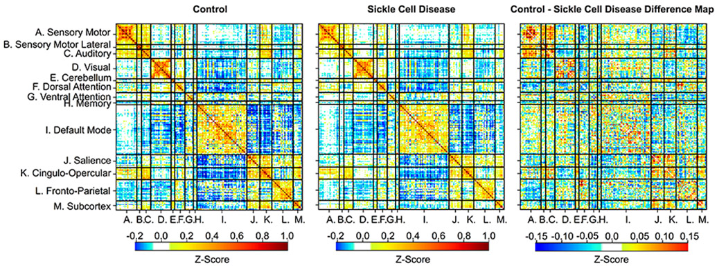
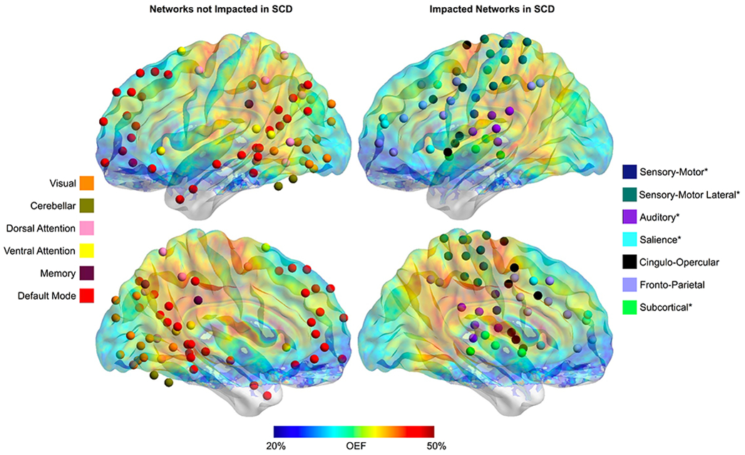
We analyzed data from 60 participants (20 controls and 40 with sickle cell disease). There was no difference in global cognition or cognitive subdomains between cohorts. However, we found decreased functional connectivity within the sensory-motor, lateral sensory-motor, auditory, salience, and subcortical networks in participants with SCD compared with controls. Further, as white matter oxygen extraction fraction increased, connectivity within the visual (p = 0.008, parameter estimate = -0.760 [95% CI = -1.297, -0.224]), default mode (p = 0.012, parameter estimate = -0.417 [95% CI = -0.731, -0.104]), and cingulo-opercular (p = 0.009, parameter estimate = -0.883 [95% CI = -1.517, -0.250]) networks decreased.
We conclude that there is diminished functional connectivity within these anatomically contiguous networks in children with SCD compared with controls, even when differences are not seen with cognitive testing. Increased white matter oxygen extraction fraction was associated with decreased connectivity in select networks. These data suggest that elevated oxygen extraction fraction and disrupted functional connectivity are potentially presymptomatic neuroimaging biomarkers for cognitive decline in SCD. ANN NEUROL 2020;88:995-1008.
即使未发生卒中,镰状细胞病(SCD)患儿也会出现认知功能缺陷。我们采用功能连接磁共振成像(fMRI)作为认知功能的潜在生物标志物,检验以下假说,即 SCD 患儿的功能连接会减少,并且氧摄取分数升高表明代谢应激最大的患儿的连接程度最低。
我们前瞻性地获取了健康对照组和 SCD 患儿的脑部 MRI 和认知测试结果。
我们分析了 60 名参与者(20 名对照组和 40 名 SCD 患儿)的数据。两组在整体认知或认知子领域方面均无差异。然而,与对照组相比,SCD 患儿的感觉运动、外侧感觉运动、听觉、突显和皮质下网络内的功能连接减少。此外,随着白质氧摄取分数增加,视觉网络(p = 0.008,参数估计值 = -0.760 [95%CI = -1.297,-0.224])、默认模式网络(p = 0.012,参数估计值 = -0.417 [95%CI = -0.731,-0.104])和扣带前回网络(p = 0.009,参数估计值 = -0.883 [95%CI = -1.517,-0.250])的连接减少。
我们的结论是,与对照组相比,即使认知测试未发现差异,SCD 患儿这些解剖上相邻网络的功能连接也会减少。白质氧摄取分数增加与特定网络的连接减少有关。这些数据表明,氧摄取分数升高和功能连接中断可能是 SCD 认知能力下降的潜在症状前神经影像学生物标志物。