College of Optometry, University of Houston, Houston, Texas, United States of America.
Children's Nutrition Research Center, Baylor College of Medicine, Houston, Texas, United States of America.
PLoS One. 2020 Sep 4;15(9):e0238750. doi: 10.1371/journal.pone.0238750. eCollection 2020.
The purpose of this study was to use a mouse model of diet-induced obesity to determine if corneal dysfunction begins prior to the onset of sustained hyperglycemia and if the dysfunction is ameliorated by diet reversal.
Six-week-old male C57BL/6 mice were fed a high fat diet (HFD) or a normal diet (ND) for 5-15 weeks. Diet reversal (DiR) mice were fed a HFD for 5 weeks, followed by a ND for 5 or 10 weeks. Corneal sensitivity was determined using aesthesiometry. Corneal cytokine expression was analyzed using a 32-plex Luminex assay. Excised corneas were prepared for immunofluorescence microscopy to evaluate diet-induced changes and wound healing. For wounding studies, mice were fed a HFD or a ND for 10 days prior to receiving a central 2mm corneal abrasion.
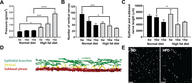
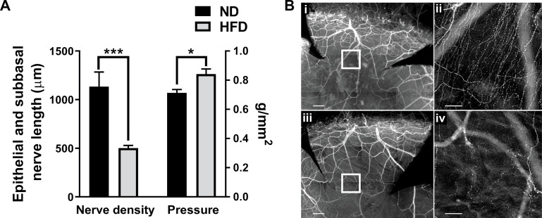
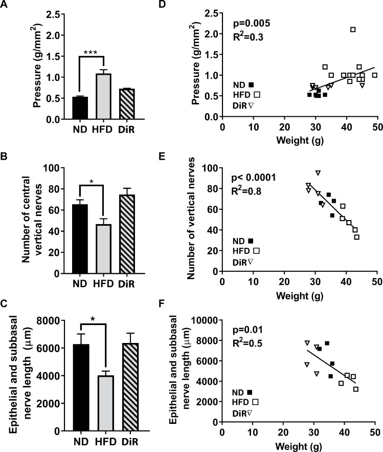
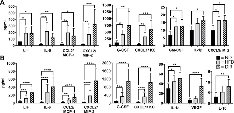
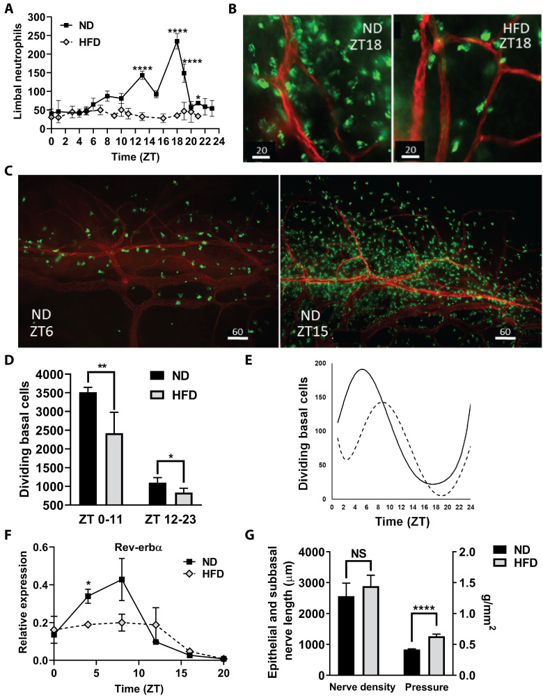
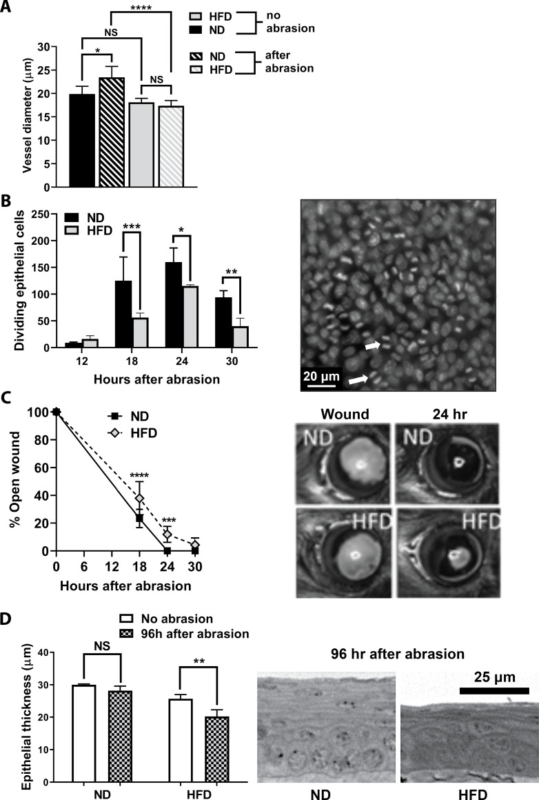
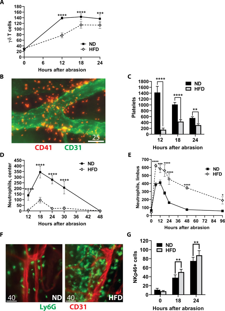
After 10 days of HFD consumption, corneal sensitivity declined. By 10 weeks, expression of corneal inflammatory mediators increased and nerve density declined. While diet reversal restored nerve density and sensitivity, the corneas remained in a heightened inflammatory state. After 10 days on the HFD, corneal circadian rhythms (limbal neutrophil accumulation, epithelial cell division and Rev-erbα expression) were blunted. Similarly, leukocyte recruitment after wounding was dysregulated and accompanied by delays in wound closure and nerve recovery.
In the mouse, obesogenic diet consumption results in corneal dysfunction that precedes the onset of sustained hyperglycemia. Diet reversal only partially ameliorated this dysfunction, suggesting a HFD diet may have a lasting negative impact on corneal health that is resistant to dietary therapeutic intervention.
本研究旨在利用饮食诱导肥胖的小鼠模型,确定角膜功能障碍是否在持续高血糖发生之前开始,以及饮食逆转是否可以改善这种功能障碍。
将 6 周龄雄性 C57BL/6 小鼠分别用高脂肪饮食(HFD)或正常饮食(ND)喂养 5-15 周。饮食逆转(DiR)组小鼠先用 HFD 喂养 5 周,然后再用 ND 喂养 5 或 10 周。采用触觉测量法测定角膜敏感性。通过 32 通道 Luminex 检测分析角膜细胞因子表达。制备离体角膜进行免疫荧光显微镜检查,以评估饮食诱导的变化和伤口愈合。对于伤口研究,在给予中央 2mm 角膜擦伤前,将小鼠用 HFD 或 ND 喂养 10 天。
HFD 喂养 10 天后,角膜敏感性下降。10 周时,角膜炎症介质的表达增加,神经密度下降。虽然饮食逆转恢复了神经密度和敏感性,但角膜仍处于高度炎症状态。HFD 喂养 10 天后,角膜昼夜节律(角膜缘中性粒细胞积聚、上皮细胞分裂和 Rev-erbα 表达)减弱。同样,创伤后白细胞募集失调,伴有伤口闭合和神经恢复延迟。
在小鼠中,肥胖饮食可导致角膜功能障碍,早于持续高血糖的发生。饮食逆转仅部分改善了这种功能障碍,表明高脂肪饮食可能对角膜健康产生持久的负面影响,且这种影响对饮食治疗干预具有抗性。