• 文献检索
  • 文档翻译
  • 深度研究
  • 学术资讯
  • Suppr Zotero 插件Zotero 插件
  • 邀请有礼
  • 套餐&价格
  • 历史记录
应用&插件
Suppr Zotero 插件Zotero 插件浏览器插件Mac 客户端Windows 客户端微信小程序
定价
高级版会员购买积分包购买API积分包
服务
文献检索文档翻译深度研究API 文档MCP 服务
关于我们
关于 Suppr公司介绍联系我们用户协议隐私条款
关注我们

Suppr 超能文献

核心技术专利:CN118964589B侵权必究
粤ICP备2023148730 号-1Suppr @ 2026

文献检索

告别复杂PubMed语法,用中文像聊天一样搜索,搜遍4000万医学文献。AI智能推荐,让科研检索更轻松。

立即免费搜索

文件翻译

保留排版,准确专业,支持PDF/Word/PPT等文件格式,支持 12+语言互译。

免费翻译文档

深度研究

AI帮你快速写综述,25分钟生成高质量综述,智能提取关键信息,辅助科研写作。

立即免费体验

乳腺弥散加权磁共振成像:图像采集和解读的标准化。

Diffusion-Weighted Magnetic Resonance Imaging of the Breast: Standardization of Image Acquisition and Interpretation.

机构信息

Department of Radiology, Seoul National University Hospital, Seoul, Korea.

Department of Radiology, Asan Medical Center, University of Ulsan College of Medicine, Seoul, Korea.

出版信息

Korean J Radiol. 2021 Jan;22(1):9-22. doi: 10.3348/kjr.2020.0093. Epub 2020 Aug 28.

DOI:10.3348/kjr.2020.0093
PMID:32901461
原文链接:https://pmc.ncbi.nlm.nih.gov/articles/PMC7772373/
Abstract

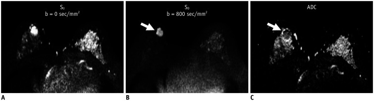
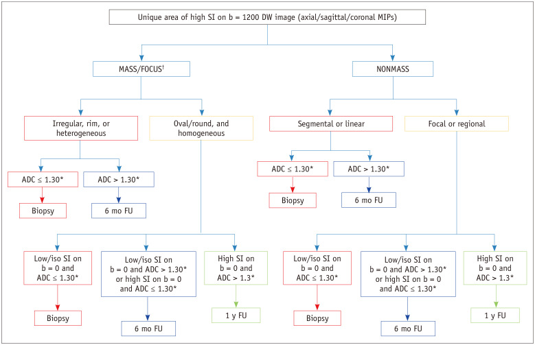
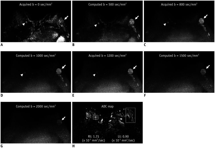
Diffusion-weighted (DW) magnetic resonance imaging (MRI) is a rapid, unenhanced imaging technique that measures the motion of water molecules within tissues and provides information regarding the cell density and tissue microstructure. DW MRI has demonstrated the potential to improve the specificity of breast MRI, facilitate the evaluation of tumor response to neoadjuvant chemotherapy and can be employed in unenhanced MRI screening. However, standardization of the acquisition and interpretation of DW MRI is challenging. Recently, the European Society of Breast Radiology issued a consensus statement, which described the acquisition parameters and interpretation of DW MRI. The current article describes the basic principles, standardized acquisition protocols and interpretation guidelines, and the clinical applications of DW MRI in breast imaging.

摘要

弥散加权(DW)磁共振成像(MRI)是一种快速、无需增强的成像技术,可测量组织内水分子的运动,提供有关细胞密度和组织微结构的信息。DW MRI 已经证明有潜力提高乳腺 MRI 的特异性,有助于评估肿瘤对新辅助化疗的反应,并且可以用于增强 MRI 筛查。然而,DW MRI 的采集和解释的标准化具有挑战性。最近,欧洲乳腺放射学会发布了一份共识声明,描述了 DW MRI 的采集参数和解读。本文介绍了 DW MRI 在乳腺成像中的基本原理、标准化采集方案和解读指南以及临床应用。

https://cdn.ncbi.nlm.nih.gov/pmc/blobs/0e68/7772373/4db2aa9734b8/kjr-22-9-g006.jpg
https://cdn.ncbi.nlm.nih.gov/pmc/blobs/0e68/7772373/e32be3938f1d/kjr-22-9-g001.jpg
https://cdn.ncbi.nlm.nih.gov/pmc/blobs/0e68/7772373/7a16c13ab85d/kjr-22-9-g002.jpg
https://cdn.ncbi.nlm.nih.gov/pmc/blobs/0e68/7772373/0e6cc5feebdd/kjr-22-9-g003.jpg
https://cdn.ncbi.nlm.nih.gov/pmc/blobs/0e68/7772373/6ad19faee9ba/kjr-22-9-g004.jpg
https://cdn.ncbi.nlm.nih.gov/pmc/blobs/0e68/7772373/fbe77decdb66/kjr-22-9-g005.jpg
https://cdn.ncbi.nlm.nih.gov/pmc/blobs/0e68/7772373/4db2aa9734b8/kjr-22-9-g006.jpg

相似文献

1
Diffusion-Weighted Magnetic Resonance Imaging of the Breast: Standardization of Image Acquisition and Interpretation.乳腺弥散加权磁共振成像:图像采集和解读的标准化。
Korean J Radiol. 2021 Jan;22(1):9-22. doi: 10.3348/kjr.2020.0093. Epub 2020 Aug 28.
2
Diffusion-weighted imaging of the breast-a consensus and mission statement from the EUSOBI International Breast Diffusion-Weighted Imaging working group.乳腺弥散加权成像的共识和任务声明:来自 EUSOBI 国际乳腺弥散加权成像工作组。
Eur Radiol. 2020 Mar;30(3):1436-1450. doi: 10.1007/s00330-019-06510-3. Epub 2019 Nov 30.
3
Diffusion-weighted MRI at 3.0 T for detection of occult disease in the contralateral breast in women with newly diagnosed breast cancer.3.0T 磁共振弥散加权成像在诊断为乳腺癌的女性对侧乳腺隐匿性疾病的检测中的应用。
Breast Cancer Res Treat. 2020 Jul;182(2):283-297. doi: 10.1007/s10549-020-05697-0. Epub 2020 May 23.
4
Diffusion-weighted MRI for Unenhanced Breast Cancer Screening.弥散加权 MRI 在乳腺癌筛查中的应用。
Radiology. 2019 Dec;293(3):504-520. doi: 10.1148/radiol.2019182789. Epub 2019 Oct 8.
5
Role of diffusion-weighted MRI: predicting axillary lymph node metastases in breast cancer.扩散加权磁共振成像的作用:预测乳腺癌腋窝淋巴结转移
Acta Radiol. 2014 Oct;55(8):909-16. doi: 10.1177/0284185113509094. Epub 2013 Nov 14.
6
Diffusion-weighted breast MRI: Clinical applications and emerging techniques.扩散加权乳腺磁共振成像:临床应用与新兴技术。
J Magn Reson Imaging. 2017 Feb;45(2):337-355. doi: 10.1002/jmri.25479. Epub 2016 Sep 30.
7
Intravoxel incoherent motion diffusion-weighted MRI for predicting response to neoadjuvant chemotherapy in breast cancer.体素内不相干运动扩散加权磁共振成像预测乳腺癌新辅助化疗疗效
Magn Reson Imaging. 2018 May;48:27-33. doi: 10.1016/j.mri.2017.12.018. Epub 2017 Dec 24.
8
Discrimination of Breast Cancer from Healthy Breast Tissue Using a Three-component Diffusion-weighted MRI Model.使用三成分扩散加权 MRI 模型鉴别乳腺癌与健康乳腺组织。
Clin Cancer Res. 2021 Feb 15;27(4):1094-1104. doi: 10.1158/1078-0432.CCR-20-2017. Epub 2020 Nov 4.
9
Differential diagnosis of benign and malignant breast masses using diffusion-weighted magnetic resonance imaging.使用扩散加权磁共振成像对乳腺良恶性肿块进行鉴别诊断。
World J Surg Oncol. 2015 Feb 7;13:32. doi: 10.1186/s12957-014-0431-3.
10
Independent value of image fusion in unenhanced breast MRI using diffusion-weighted and morphological T2-weighted images for lesion characterization in patients with recently detected BI-RADS 4/5 x-ray mammography findings.在未增强乳腺MRI中,利用扩散加权成像和形态学T2加权成像进行图像融合,对近期乳腺影像报告和数据系统(BI-RADS)4/5级乳腺钼靶X线摄影检查结果的患者进行病变特征分析的独立价值。
Eur Radiol. 2017 Feb;27(2):562-569. doi: 10.1007/s00330-016-4400-9. Epub 2016 May 18.

引用本文的文献

1
Breast Cancer Screening in Asian Countries: Epidemiology, Screening Practices, Outcomes, Challenges, and Future Directions.亚洲国家的乳腺癌筛查:流行病学、筛查实践、结果、挑战及未来方向
Korean J Radiol. 2025 Aug;26(8):743-758. doi: 10.3348/kjr.2025.0338.
2
Preoperative Diagnosis of Ipsilateral and Contralateral Breast Cancer: Role of Diffusion-weighted MRI.同侧及对侧乳腺癌的术前诊断:扩散加权磁共振成像的作用
Radiology. 2025 Jun;315(3):e242423. doi: 10.1148/radiol.242423.
3
Magnetic resonance imaging characteristics and survival outcomes of metaplastic breast carcinoma.

本文引用的文献

1
Diffusion-weighted imaging of the breast-a consensus and mission statement from the EUSOBI International Breast Diffusion-Weighted Imaging working group.乳腺弥散加权成像的共识和任务声明:来自 EUSOBI 国际乳腺弥散加权成像工作组。
Eur Radiol. 2020 Mar;30(3):1436-1450. doi: 10.1007/s00330-019-06510-3. Epub 2019 Nov 30.
2
Diffusion-weighted MRI for Unenhanced Breast Cancer Screening.弥散加权 MRI 在乳腺癌筛查中的应用。
Radiology. 2019 Dec;293(3):504-520. doi: 10.1148/radiol.2019182789. Epub 2019 Oct 8.
3
Diffusion tensor imaging for characterizing tumor microstructure and improving diagnostic performance on breast MRI: a prospective observational study.
化生性乳腺癌的磁共振成像特征及生存结果
PLoS One. 2025 May 14;20(5):e0323541. doi: 10.1371/journal.pone.0323541. eCollection 2025.
4
Dynamic contrast-enhanced magnetic resonance imaging parameters combined with diffusion-weighted imaging for discriminating malignant lesions, molecular subtypes, and pathological grades in invasive ductal carcinoma patients.动态对比增强磁共振成像参数联合扩散加权成像用于鉴别浸润性导管癌患者的恶性病变、分子亚型和病理分级
PLoS One. 2025 Apr 15;20(4):e0320240. doi: 10.1371/journal.pone.0320240. eCollection 2025.
5
Breast Diffusion-weighted MR Imaging: Current Applications, Insights from Screening, and Future Directions.乳腺扩散加权磁共振成像:当前应用、筛查见解及未来方向。
Magn Reson Med Sci. 2025 Jul 1;24(3):300-314. doi: 10.2463/mrms.rev.2024-0142. Epub 2025 Feb 8.
6
Performance of high-resolution diffusion-weighted magnetic resonance imaging for detecting clinically occult early breast cancers: a multi-reader study.高分辨率扩散加权磁共振成像在检测临床隐匿性早期乳腺癌中的性能:一项多阅片者研究
Breast Cancer Res Treat. 2025 Feb;210(1):71-86. doi: 10.1007/s10549-024-07537-x. Epub 2024 Nov 8.
7
Fat-signal suppression in breast diffusion-weighted imaging: the Good, the Bad, and the Ugly.乳腺扩散加权成像中的脂肪信号抑制:优点、缺点与问题
Eur Radiol. 2025 Feb;35(2):733-741. doi: 10.1007/s00330-024-10973-4. Epub 2024 Aug 7.
8
Diffusion-Weighted MRI for the Assessment of Molecular Prognostic Biomarkers in Breast Cancer.扩散加权磁共振成像在乳腺癌分子预后生物标志物评估中的应用。
Korean J Radiol. 2024 Jul;25(7):623-633. doi: 10.3348/kjr.2023.1188.
9
Background Breast Parenchymal Signal During Menstrual Cycle on Diffusion-Weighted MRI: A Prospective Study in Healthy Premenopausal Women.背景:月经周期中乳腺实质信号在弥散加权 MRI 中的表现:一项对健康绝经前女性的前瞻性研究。
Korean J Radiol. 2024 Jun;25(6):511-517. doi: 10.3348/kjr.2023.1189.
10
Non-contrast Breast MR Imaging.乳腺非增强磁共振成像
Radiol Clin North Am. 2024 Jul;62(4):661-678. doi: 10.1016/j.rcl.2023.12.009. Epub 2024 Mar 7.
弥散张量成像用于表征肿瘤微结构并提高乳腺 MRI 的诊断性能:一项前瞻性观察研究。
Breast Cancer Res. 2019 Sep 4;21(1):102. doi: 10.1186/s13058-019-1183-3.
4
Utility of Diffusion-weighted Imaging to Decrease Unnecessary Biopsies Prompted by Breast MRI: A Trial of the ECOG-ACRIN Cancer Research Group (A6702).扩散加权成像在减少乳腺 MRI 引导的不必要活检中的应用:ECOG-ACRIN 癌症研究组(A6702)的一项试验。
Clin Cancer Res. 2019 Mar 15;25(6):1756-1765. doi: 10.1158/1078-0432.CCR-18-2967. Epub 2019 Jan 15.
5
Evaluation of Breast Cancer Morphology Using Diffusion-Weighted and Dynamic Contrast-Enhanced MRI: Intermethod and Interobserver Agreement.使用扩散加权和动态对比增强 MRI 评估乳腺癌形态:方法间和观察者间的一致性。
J Magn Reson Imaging. 2019 May;49(5):1381-1390. doi: 10.1002/jmri.26332. Epub 2018 Oct 16.
6
Intravoxel incoherent motion diffusion-weighted MRI of invasive breast cancer: Correlation with prognostic factors and kinetic features acquired with computer-aided diagnosis.浸润性乳腺癌体素内不相干运动扩散加权 MRI:与预后因素及计算机辅助诊断获得的动力学特征的相关性。
J Magn Reson Imaging. 2019 Jan;49(1):118-130. doi: 10.1002/jmri.26221. Epub 2018 Sep 20.
7
Diffusion-weighted MRI Findings Predict Pathologic Response in Neoadjuvant Treatment of Breast Cancer: The ACRIN 6698 Multicenter Trial.扩散加权 MRI 表现预测乳腺癌新辅助治疗的病理反应:ACRIN 6698 多中心试验。
Radiology. 2018 Dec;289(3):618-627. doi: 10.1148/radiol.2018180273. Epub 2018 Sep 4.
8
Diffusion tensor magnetic resonance imaging of breast cancer: associations between diffusion metrics and histological prognostic factors.乳腺癌的弥散张量磁共振成像:弥散指标与组织学预后因素的相关性。
Eur Radiol. 2018 Aug;28(8):3185-3193. doi: 10.1007/s00330-018-5429-8. Epub 2018 Apr 30.
9
Diffusion-Weighted Imaging With Apparent Diffusion Coefficient Mapping for Breast Cancer Detection as a Stand-Alone Parameter: Comparison With Dynamic Contrast-Enhanced and Multiparametric Magnetic Resonance Imaging.扩散加权成像联合表观扩散系数图在乳腺癌检测中的应用:与动态对比增强和多参数磁共振成像的比较。
Invest Radiol. 2018 Oct;53(10):587-595. doi: 10.1097/RLI.0000000000000465.
10
Potential of Noncontrast Magnetic Resonance Imaging With Diffusion-Weighted Imaging in Characterization of Breast Lesions: Intraindividual Comparison With Dynamic Contrast-Enhanced Magnetic Resonance Imaging.弥散加权成像联合非增强磁共振成像在乳腺病变定性诊断中的价值:与动态对比增强磁共振成像的个体内对照研究。
Invest Radiol. 2018 Apr;53(4):229-235. doi: 10.1097/RLI.0000000000000433.