Kim Taeho, Lewis Benjamin C, Price Alex, Mazur Thomas, Gach H Michael, Park Justin C, Cai Bin, Wittland Erin, Henke Lauren, Kim Hyun, Mutic Sasa, Green Olga
Department of Radiation Oncology, Washington University School of Medicine, St Louis, MO, 63110, USA.
Department of Radiology and Biomedical Engineering, Washington University in St. Louis, St Louis, MO, 63110, USA.
J Appl Clin Med Phys. 2020 Oct;21(10):241-247. doi: 10.1002/acm2.13016. Epub 2020 Sep 15.
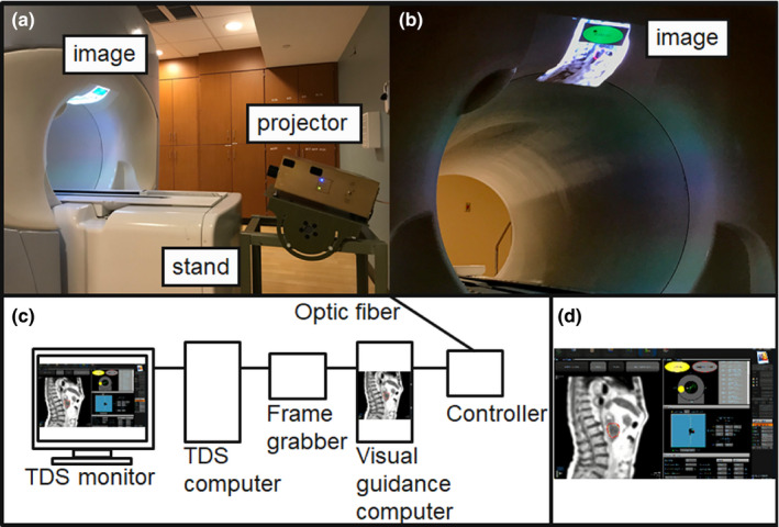
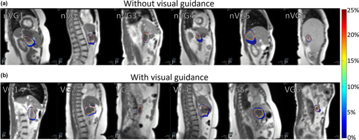
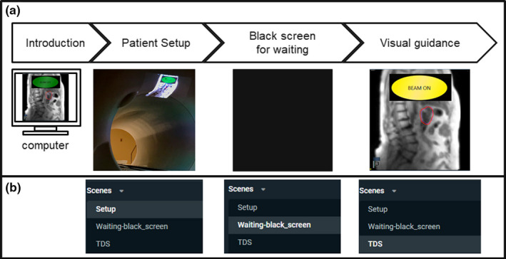
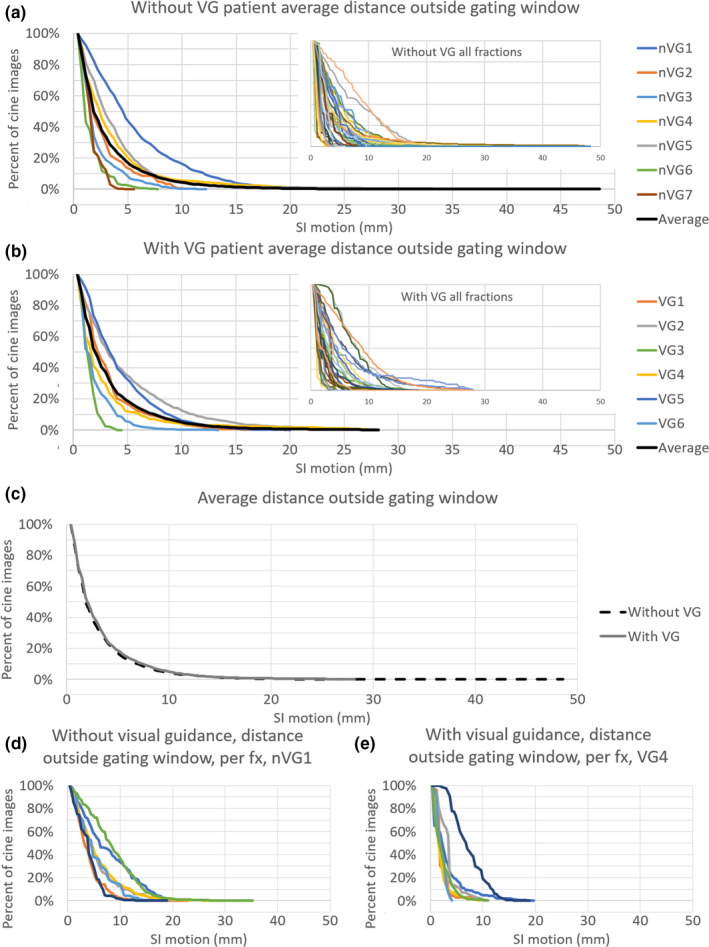
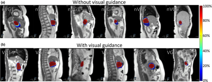
To present a tumor motion control system during free breathing using direct tumor visual feedback to patients in 0.35 T magnetic resonance-guided radiotherapy (MRgRT). We present direct tumor visualization to patients by projecting real-time cine MR images on an MR-compatible display system inside a 0.35 T MRgRT bore. The direct tumor visualization included anatomical images with a target contour and an auto-segmented gating contour. In addition, a beam-status sign was added for patient guidance. The feasibility was investigated with a six-patient clinical evaluation of the system in terms of tumor motion range and beam-on time. Seven patients without visual guidance were used for comparison. Positions of the tumor and the auto-segmented gating contour from the cine MR images were used in probability analysis to evaluate tumor motion control. In addition, beam-on time was recorded to assess the efficacy of the visual feedback system. The direct tumor visualization system was developed and implemented in our clinic. The target contour extended 3 mm outside of the gating contour for 33.6 ± 24.9% of the time without visual guidance, and 37.2 ± 26.4% of the time with visual guidance. The average maximum motion outside of the gating contour was 14.4 ± 11.1 mm without and 13.0 ± 7.9 mm with visual guidance. Beam-on time as a percentage was 43.9 ± 15.3% without visual guidance, and 48.0 ± 21.2% with visual guidance, but was not significantly different (P = 0.34). We demonstrated the clinical feasibility and potential benefits of presenting direct tumor visual feedback to patients in MRgRT. The visual feedback allows patients to visualize and attempt to minimize tumor motion in free breathing. The proposed system and associated clinical workflow can be easily adapted for any type of MRgRT.
在0.35T磁共振引导放疗(MRgRT)中,利用直接肿瘤视觉反馈向患者呈现自由呼吸期间的肿瘤运动控制系统。我们通过将实时电影磁共振图像投影到0.35T MRgRT孔径内的磁共振兼容显示系统上,向患者呈现直接肿瘤可视化。直接肿瘤可视化包括带有目标轮廓和自动分割门控轮廓的解剖图像。此外,还添加了光束状态标志以指导患者。通过对该系统进行六例患者的临床评估,从肿瘤运动范围和照射时间方面研究了其可行性。七例没有视觉引导的患者用于比较。电影磁共振图像中肿瘤和自动分割门控轮廓的位置用于概率分析,以评估肿瘤运动控制。此外,记录照射时间以评估视觉反馈系统的效果。直接肿瘤可视化系统已在我们的诊所开发并实施。在没有视觉引导的情况下,目标轮廓在门控轮廓外延伸3mm的时间占33.6±24.9%,有视觉引导时占37.2±26.4%。在门控轮廓外的平均最大运动,没有视觉引导时为14.4±11.1mm,有视觉引导时为13.0±7.9mm。照射时间百分比在没有视觉引导时为43.9±15.3%,有视觉引导时为48.0±21.2%,但差异不显著(P = 0.34)。我们证明了在MRgRT中向患者呈现直接肿瘤视觉反馈的临床可行性和潜在益处。视觉反馈使患者能够在自由呼吸时可视化并试图最小化肿瘤运动。所提出的系统和相关临床工作流程可轻松适用于任何类型的MRgRT。