Strutural Biology of Disease Processes Laboratory, Francis Crick Institute, London, UK.
Precision Medicine Center, The Seventh Affiliated Hospital, Sun Yat-sen University, Shenzhen, China.
Nature. 2020 Dec;588(7837):327-330. doi: 10.1038/s41586-020-2772-0. Epub 2020 Sep 17.
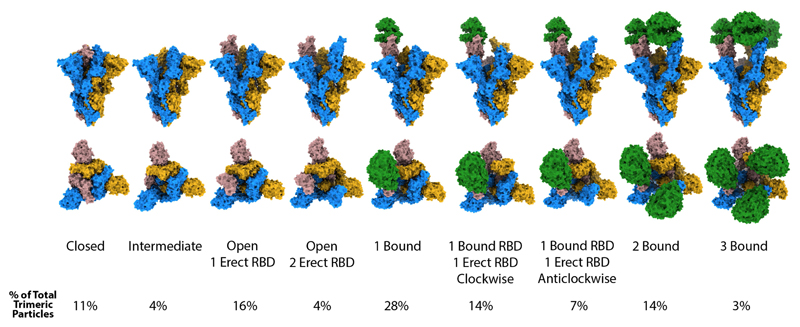
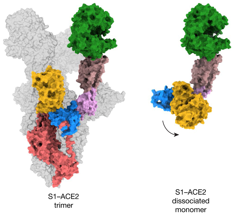
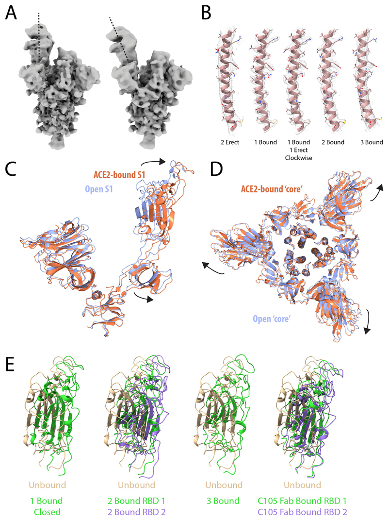
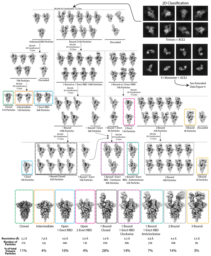
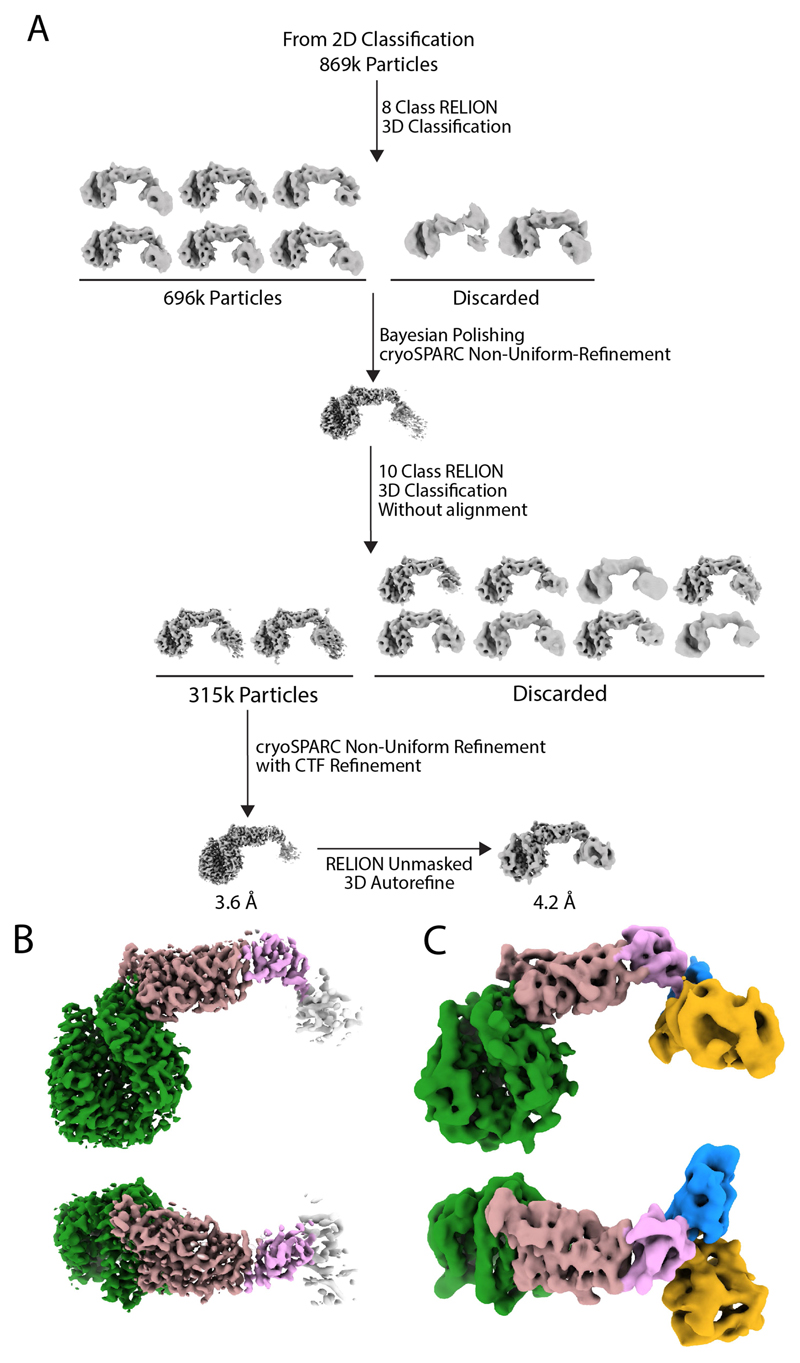
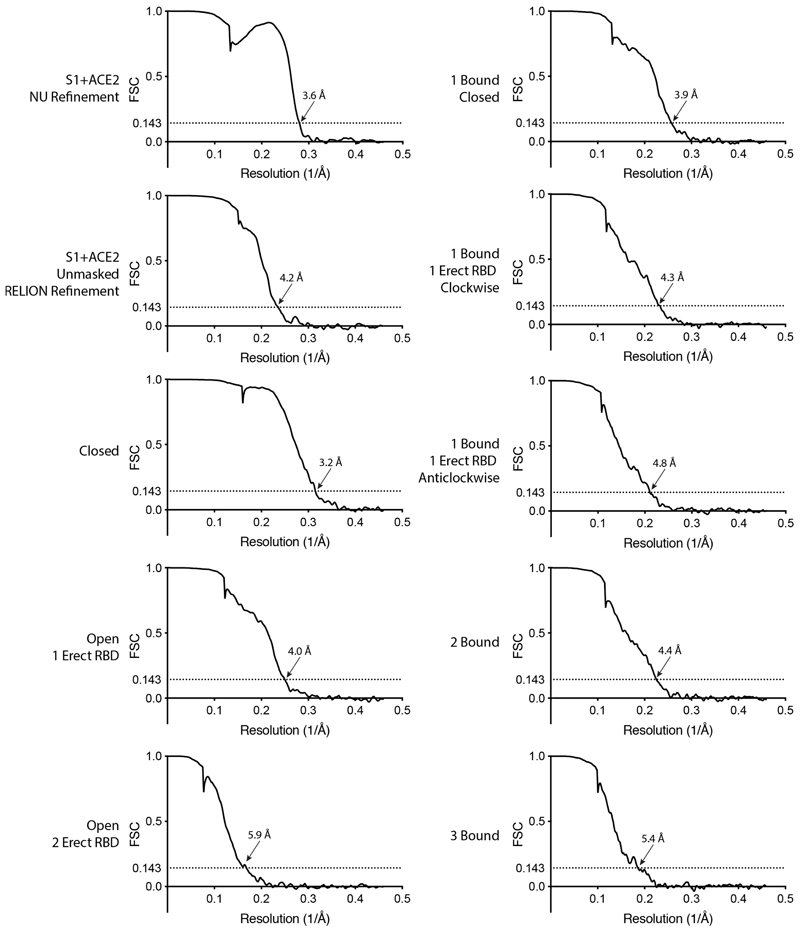
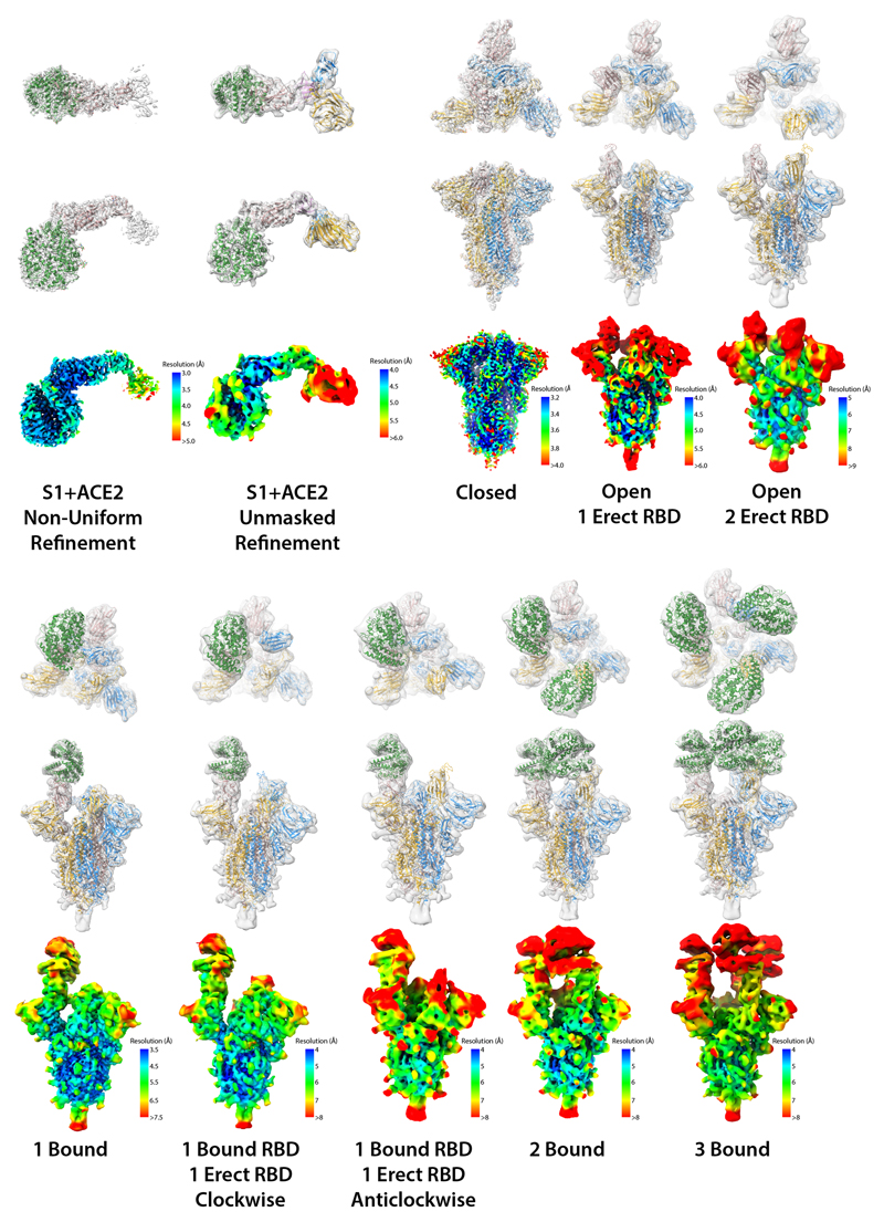
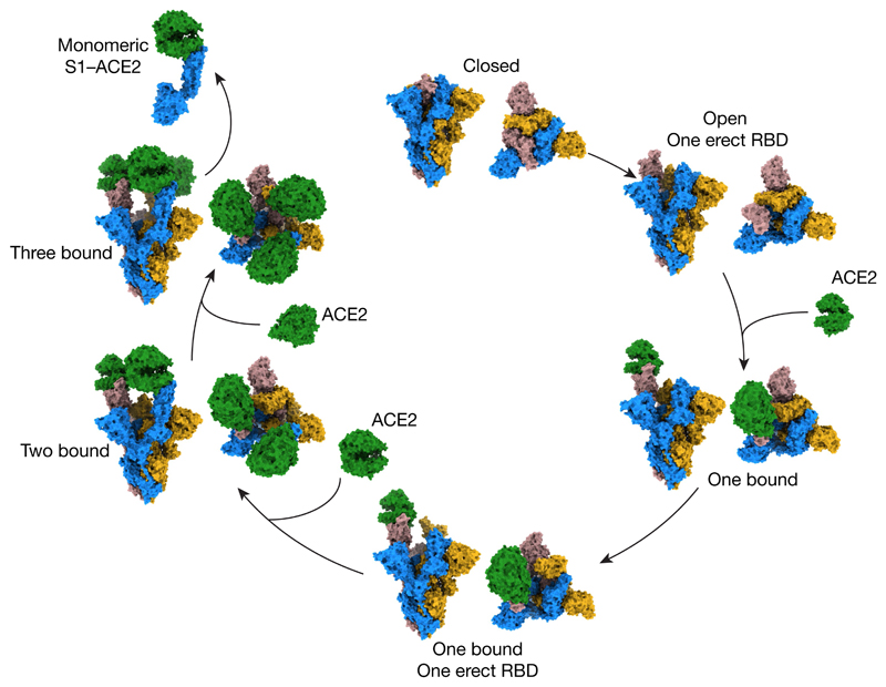
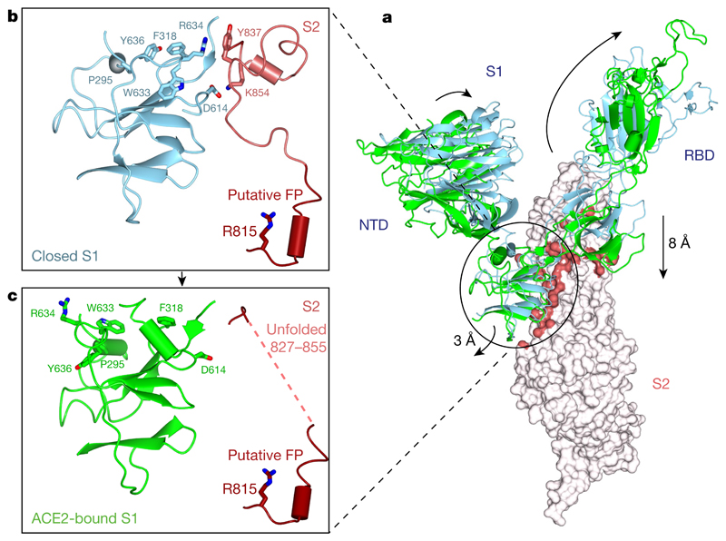
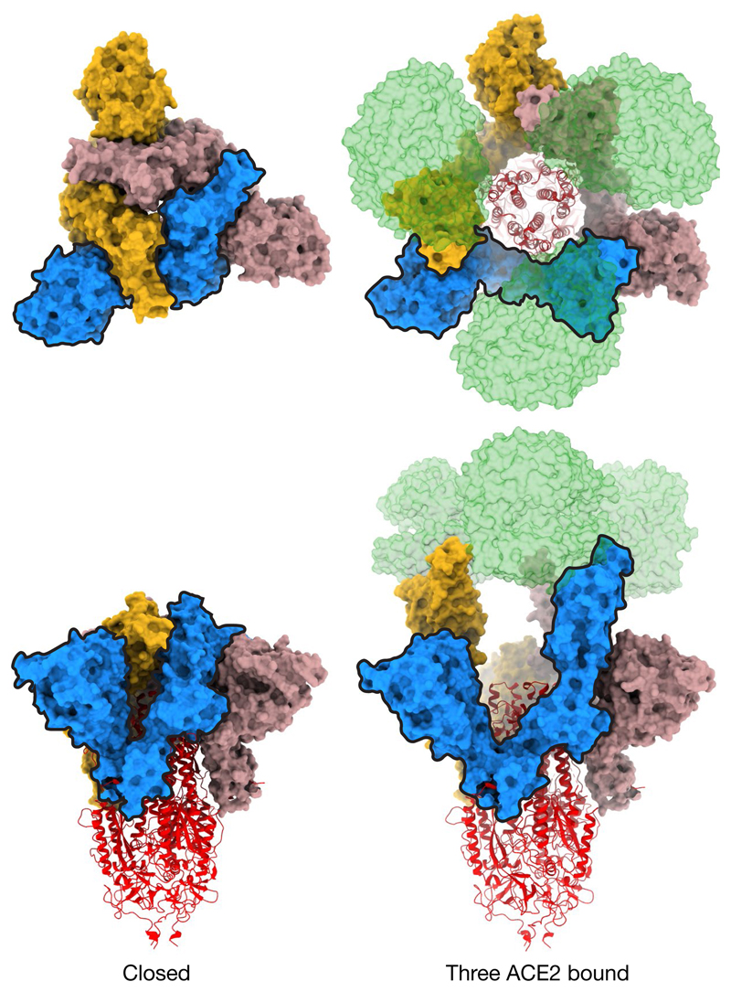
Infection with severe acute respiratory syndrome coronavirus 2 (SARS-CoV-2) is initiated by virus binding to the ACE2 cell-surface receptors, followed by fusion of the virus and cell membranes to release the virus genome into the cell. Both receptor binding and membrane fusion activities are mediated by the virus spike glycoprotein. As with other class-I membrane-fusion proteins, the spike protein is post-translationally cleaved, in this case by furin, into the S1 and S2 components that remain associated after cleavage. Fusion activation after receptor binding is proposed to involve the exposure of a second proteolytic site (S2'), cleavage of which is required for the release of the fusion peptide. Here we analyse the binding of ACE2 to the furin-cleaved form of the SARS-CoV-2 spike protein using cryo-electron microscopy. We classify ten different molecular species, including the unbound, closed spike trimer, the fully open ACE2-bound trimer and dissociated monomeric S1 bound to ACE2. The ten structures describe ACE2-binding events that destabilize the spike trimer, progressively opening up, and out, the individual S1 components. The opening process reduces S1 contacts and unshields the trimeric S2 core, priming the protein for fusion activation and dissociation of ACE2-bound S1 monomers. The structures also reveal refolding of an S1 subdomain after ACE2 binding that disrupts interactions with S2, which involves Asp614 and leads to the destabilization of the structure of S2 proximal to the secondary (S2') cleavage site.
严重急性呼吸综合征冠状病毒 2 (SARS-CoV-2) 的感染是由病毒与 ACE2 细胞表面受体结合引发的,随后病毒和细胞膜融合将病毒基因组释放到细胞内。受体结合和膜融合活性均由病毒刺突糖蛋白介导。与其他 I 类膜融合蛋白一样,刺突蛋白在翻译后被弗林蛋白酶切割,在这种情况下切割成 S1 和 S2 两个组成部分,切割后仍保持关联。受体结合后的融合激活被认为涉及第二个蛋白水解位点 (S2') 的暴露,该位点的切割对于融合肽的释放是必需的。在这里,我们使用冷冻电子显微镜分析 ACE2 与 SARS-CoV-2 刺突蛋白的弗林蛋白酶切割形式的结合。我们将十种不同的分子物种进行分类,包括未结合的、封闭的刺突三聚体、完全开放的 ACE2 结合的三聚体和与 ACE2 结合的分离的单体 S1。这十个结构描述了 ACE2 结合事件,这些事件使刺突三聚体不稳定,逐渐打开并向外展开各个 S1 成分。打开过程减少了 S1 的接触并使三聚体 S2 核心暴露,为融合激活和 ACE2 结合的 S1 单体的解离做好准备。这些结构还揭示了 ACE2 结合后 S1 亚结构域的重折叠,该重折叠破坏了与 S2 的相互作用,涉及 Asp614,并导致靠近二级 (S2') 切割位点的 S2 近端结构的不稳定。