Csutak Csaba, Ștefan Paul-Andrei, Lenghel Lavinia Manuela, Moroșanu Cezar Octavian, Lupean Roxana-Adelina, Șimonca Larisa, Mihu Carmen Mihaela, Lebovici Andrei
Radiology and Imaging Department, County Emergency Hospital, Cluj-Napoca, Clinicilor Street, Number 5, Cluj-Napoca, 400006 Cluj, Romania.
Radiology, Surgical Specialties Department, "Iuliu Haţieganu" University of Medicine and Pharmacy, Clinicilor Street, number 3-5, Cluj-Napoca, 400006 Cluj, Romania.
Brain Sci. 2020 Sep 16;10(9):638. doi: 10.3390/brainsci10090638.
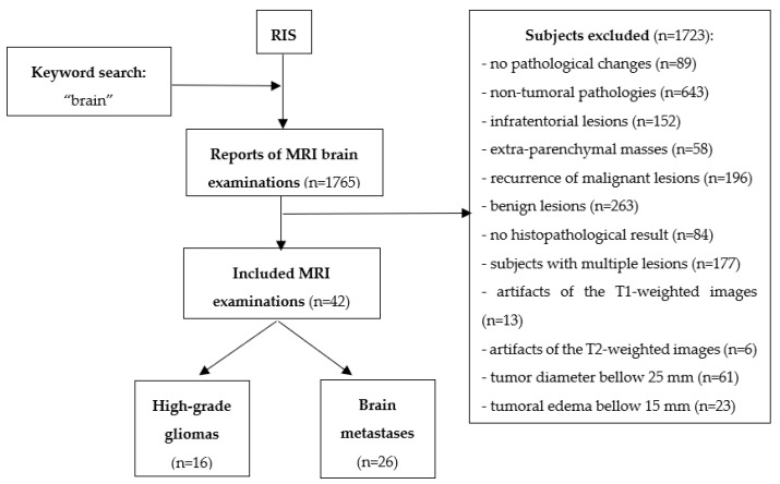
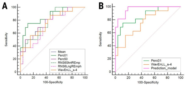
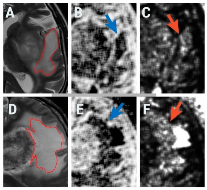
High-grade gliomas (HGGs) and solitary brain metastases (BMs) have similar imaging appearances, which often leads to misclassification. In HGGs, the surrounding tissues show malignant invasion, while BMs tend to displace the adjacent area. The surrounding edema produced by the two cannot be differentiated by conventional magnetic resonance (MRI) examinations. Forty-two patients with pathology-proven brain tumors who underwent conventional pretreatment MRIs were retrospectively included (HGGs, = 16; BMs, = 26). Texture analysis of the peritumoral zone was performed on the T2-weighted sequence using dedicated software. The most discriminative texture features were selected using the Fisher and the probability of classification error and average correlation coefficients. The ability of texture parameters to distinguish between HGGs and BMs was evaluated through univariate, receiver operating, and multivariate analyses. The first percentile and wavelet energy texture parameters were independent predictors of HGGs (75-87.5% sensitivity, 53.85-88.46% specificity). The prediction model consisting of all parameters that showed statistically significant results at the univariate analysis was able to identify HGGs with 100% sensitivity and 66.7% specificity. Texture analysis can provide a quantitative description of the peritumoral zone encountered in solitary brain tumors, that can provide adequate differentiation between HGGs and BMs.
高级别胶质瘤(HGGs)和孤立性脑转移瘤(BMs)具有相似的影像学表现,这常常导致误诊。在高级别胶质瘤中,周围组织显示恶性浸润,而脑转移瘤往往使相邻区域移位。两者产生的周围水肿通过传统磁共振成像(MRI)检查无法区分。回顾性纳入42例经病理证实的脑肿瘤患者,这些患者均接受了传统的预处理MRI检查(高级别胶质瘤,n = 16;脑转移瘤,n = 26)。使用专用软件在T2加权序列上对瘤周区域进行纹理分析。使用Fisher法以及分类错误概率和平均相关系数选择最具鉴别力的纹理特征。通过单变量分析、受试者工作特征分析和多变量分析评估纹理参数区分高级别胶质瘤和脑转移瘤的能力。第一百分位数和小波能量纹理参数是高级别胶质瘤的独立预测因子(敏感性为75 - 87.5%,特异性为53.85 - 88.46%)。由单变量分析中显示具有统计学显著结果的所有参数组成的预测模型能够以100%的敏感性和66.7%的特异性识别高级别胶质瘤。纹理分析可以对孤立性脑肿瘤的瘤周区域进行定量描述,从而能够充分区分高级别胶质瘤和脑转移瘤。