• 文献检索
  • 文档翻译
  • 深度研究
  • 学术资讯
  • Suppr Zotero 插件Zotero 插件
  • 邀请有礼
  • 套餐&价格
  • 历史记录
应用&插件
Suppr Zotero 插件Zotero 插件浏览器插件Mac 客户端Windows 客户端微信小程序
定价
高级版会员购买积分包购买API积分包
服务
文献检索文档翻译深度研究API 文档MCP 服务
关于我们
关于 Suppr公司介绍联系我们用户协议隐私条款
关注我们

Suppr 超能文献

核心技术专利:CN118964589B侵权必究
粤ICP备2023148730 号-1Suppr @ 2026

文献检索

告别复杂PubMed语法,用中文像聊天一样搜索,搜遍4000万医学文献。AI智能推荐,让科研检索更轻松。

立即免费搜索

文件翻译

保留排版,准确专业,支持PDF/Word/PPT等文件格式,支持 12+语言互译。

免费翻译文档

深度研究

AI帮你快速写综述,25分钟生成高质量综述,智能提取关键信息,辅助科研写作。

立即免费体验

使用光片显微镜对成年秀丽隐杆线虫进行活体成像。

Imaging adult C. elegans live using light-sheet microscopy.

机构信息

LaserLaB and Department of Physics and Astronomy, Vrije Universiteit Amsterdam, Amsterdam, The Netherlands.

出版信息

J Microsc. 2021 Mar;281(3):214-223. doi: 10.1111/jmi.12964. Epub 2020 Oct 10.

DOI:10.1111/jmi.12964
PMID:32949409
原文链接:https://pmc.ncbi.nlm.nih.gov/articles/PMC7891351/
Abstract

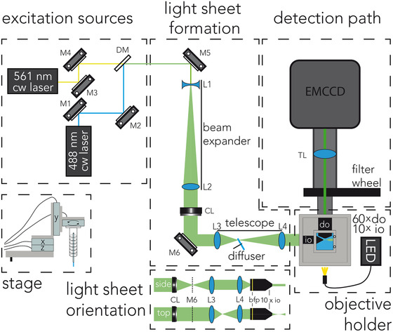
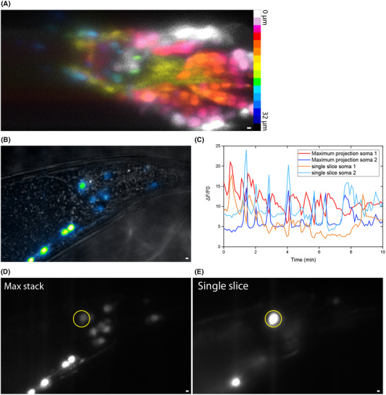
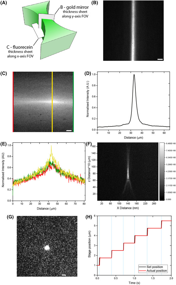
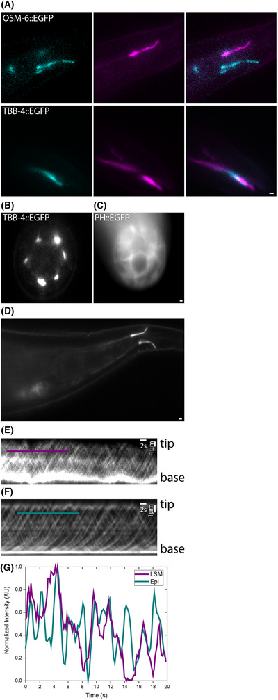
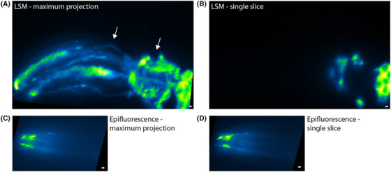
Live observation of biological phenomena in the context of living organisms can provide important insights in the mechanisms of these phenomena. However, the spatially complex and dynamic physiology of multicellular organisms can be a challenging environment to make observations with fluorescence microscopy. Due to the illumination of out-of-focus planes, confocal and particularly widefield fluorescence microscopy suffer from low signal-to-background ratio (SBR), photo toxicity and bleaching of fluorescent probes. In light-sheet microscopy (LSM), solely the focal plane of the detection objective is illuminated, minimising out-of-focus fluorescence and photobleaching, thereby enhancing SBR, allowing for low laser intensities and longer acquisition periods. Here we present a straightforward light-sheet microscope with a 1.0-NA detection objective and a fast sample-positioning stage that allows for four degrees of freedom. By imaging the sensory cilia and nervous system of living young adult C. elegans, we demonstrate that the instrument is well suited for relatively fast, volumetric imaging of larger (hundreds of micrometres cubed) living samples. These experiments demonstrate that such an instrument provides a valuable addition to commonly used widefield and confocal fluorescence microscopes. LAY DESCRIPTION: In fluorescence microscopy, sharp images can only be obtained when the light obtained from the section of the image that is in focus is not overwhelmed by light emerging from elsewhere. In this paper, we present a light-sheet fluorescence microscope, based on the OpenSPIM initiative, with a magnification of 90× and a sensitive sample positioning stage that allows for fast controlled linear movement and rotation. In a light-sheet microscope (LSM), the sample is illuminated from the side, compared to the direction of detection, limiting illumination only to the part of the sample that is imaged in the focal plane (general resources: Wikipedia or MicroscopyU). This does not only limit background noise, but also reduces damage to the sample due to phototoxicity. This makes a LSM particularly suitable for imaging living samples at high resolution, in three dimensions, over long periods of time. Our instrument was specifically designed for imaging adult C. elegans nematodes. We show here how the instrument compares to a standard epifluorescence microscope, imaging neuronal structures in the animals. The instrument proved well suited for fast volumetric imaging of larger cellular structures such as C. elegans neuronal cell bodies. Our experiments show that the instrument provides a valuable addition to widefield and confocal fluorescence microscopes commonly used to image adult C. elegans.

摘要

在活生物体的背景下对生物现象进行实时观察,可以深入了解这些现象的机制。然而,多细胞生物体的空间复杂且动态的生理学特性使得使用荧光显微镜进行观察具有挑战性。由于离焦平面的照明,共聚焦和宽场荧光显微镜的信号背景比(SBR)低,对荧光探针有光毒性和漂白作用。在光片显微镜(LSM)中,仅检测物镜的焦平面被照明,从而最小化离焦荧光和漂白,从而提高 SBR,允许使用低激光强度和更长的采集时间。在这里,我们介绍了一种具有 1.0-NA 检测物镜和快速样品定位台的简单光片显微镜,该定位台允许四个自由度。通过对活体年轻成年秀丽隐杆线虫的感觉纤毛和神经系统进行成像,我们证明该仪器非常适合对较大(数百立方微米)的活体样本进行相对快速的体积成像。这些实验表明,这种仪器是常用的宽场和共聚焦荧光显微镜的有价值的补充。

通俗描述

在荧光显微镜中,只有当从聚焦部分获取的光不被来自其他地方的光淹没时,才能获得清晰的图像。在本文中,我们提出了一种基于 OpenSPIM 计划的光片荧光显微镜,放大倍数为 90×,并且具有灵敏的样品定位台,可以进行快速的受控线性运动和旋转。在光片显微镜(LSM)中,与检测方向相比,从侧面对样品进行照明,仅将照明限制在聚焦平面中成像的样品部分(一般资源:维基百科或显微镜 U)。这不仅限制了背景噪声,而且还减少了由于光毒性对样品的损害。这使得 LSM 特别适合于在高分辨率,三维空间中长时间对活样本进行成像。我们的仪器是专门为成像成年秀丽隐杆线虫线虫而设计的。在这里,我们展示了该仪器如何与标准的落射荧光显微镜相比,对动物的神经元结构进行成像。该仪器非常适合对较大的细胞结构(例如秀丽隐杆线虫神经元胞体)进行快速体积成像。我们的实验表明,该仪器是常用的成像成年秀丽隐杆线虫的宽场和共聚焦荧光显微镜的有价值的补充。

https://cdn.ncbi.nlm.nih.gov/pmc/blobs/7787/7891351/dd5eabee3c85/JMI-281-214-g005.jpg
https://cdn.ncbi.nlm.nih.gov/pmc/blobs/7787/7891351/5a2806e5c262/JMI-281-214-g001.jpg
https://cdn.ncbi.nlm.nih.gov/pmc/blobs/7787/7891351/7146be0b1254/JMI-281-214-g002.jpg
https://cdn.ncbi.nlm.nih.gov/pmc/blobs/7787/7891351/820d989d2f37/JMI-281-214-g003.jpg
https://cdn.ncbi.nlm.nih.gov/pmc/blobs/7787/7891351/3df718362a81/JMI-281-214-g004.jpg
https://cdn.ncbi.nlm.nih.gov/pmc/blobs/7787/7891351/dd5eabee3c85/JMI-281-214-g005.jpg

相似文献

1
Imaging adult C. elegans live using light-sheet microscopy.使用光片显微镜对成年秀丽隐杆线虫进行活体成像。
J Microsc. 2021 Mar;281(3):214-223. doi: 10.1111/jmi.12964. Epub 2020 Oct 10.
2
Light-sheet microscopy for everyone? Experience of building an OpenSPIM to study flatworm development.人人都能使用的光片显微镜?构建用于研究涡虫发育的开放式选择性平面照明显微镜的经验。
BMC Dev Biol. 2016 Jun 30;16(1):22. doi: 10.1186/s12861-016-0122-0.
3
Three-dimensional Fluorescence Lifetime Imaging with a Single Plane Illumination Microscope provides an improved signal to noise ratio.使用单平面照明显微镜的三维荧光寿命成像可提供更高的信噪比。
Opt Express. 2011 Oct 10;19(21):20743-50. doi: 10.1364/OE.19.020743.
4
Confocal imaging capacity on a widefield microscope using a spatial light modulator.利用空间光调制器在宽场显微镜上实现共焦成像。
PLoS One. 2021 Feb 16;16(2):e0244034. doi: 10.1371/journal.pone.0244034. eCollection 2021.
5
Low-cost optofluidic add-on enables rapid selective plane illumination microscopy of C. elegans with a conventional wide-field microscope.低成本光流附加装置使传统宽场显微镜能够快速选择性平面照明显微观察秀丽隐杆线虫。
J Biomed Opt. 2021 Dec;26(12). doi: 10.1117/1.JBO.26.12.126501.
6
Video-rate scanning confocal microscopy and microendoscopy.视频速率扫描共聚焦显微镜和微型内窥镜检查。
J Vis Exp. 2011 Oct 20(56):3252. doi: 10.3791/3252.
7
Efficient and cost-effective 3D cellular imaging by sub-voxel-resolving light-sheet add-on microscopy.亚像素解析度的光片附加显微镜实现高效、经济的 3D 细胞成像。
J Biophotonics. 2020 Jun;13(6):e201960243. doi: 10.1002/jbio.201960243. Epub 2020 Feb 27.
8
Resolution doubling in light-sheet microscopy via oblique plane structured illumination.利用斜平面结构照明实现光片显微镜的分辨率倍增。
Nat Methods. 2022 Nov;19(11):1419-1426. doi: 10.1038/s41592-022-01635-8. Epub 2022 Oct 24.
9
Light sheet fluorescence microscopy: a review.光片荧光显微镜:综述。
J Histochem Cytochem. 2011 Feb;59(2):129-38. doi: 10.1369/0022155410394857.
10
Basic building units and properties of a fluorescence single plane illumination microscope.荧光单平面照明显微镜的基本构建单元及特性
Rev Sci Instrum. 2007 Feb;78(2):023705. doi: 10.1063/1.2428277.

引用本文的文献

1
Brief guide to Caenorhabditis elegans imaging and quantification.秀丽隐杆线虫成像与定量简要指南。
Mol Cells. 2025 Jun 29;48(9):100249. doi: 10.1016/j.mocell.2025.100249.
2
Enhanced neuroimaging with a calcium sensor in brains using closed-loop adaptive optics light-sheet fluorescence microscopy.利用闭环自适应光学光片荧光显微镜在大脑中使用钙传感器进行增强的神经影像学研究。
J Biomed Opt. 2023 Jun;28(6):066501. doi: 10.1117/1.JBO.28.6.066501. Epub 2023 Jun 16.
3
Single-molecule imaging in the primary cilium.初级纤毛中单分子成像。

本文引用的文献

1
A parthenogenetic quasi-program causes teratoma-like tumors during aging in wild-type .一种孤雌生殖准程序在野生型衰老过程中引发畸胎瘤样肿瘤。
NPJ Aging Mech Dis. 2018 Jun 13;4:6. doi: 10.1038/s41514-018-0025-3. eCollection 2018.
2
An improved inverse-type Ca2+ indicator can detect putative neuronal inhibition in Caenorhabditis elegans by increasing signal intensity upon Ca2+ decrease.一种改良的反向型 Ca2+ 指示剂可以通过在 Ca2+ 减少时增加信号强度来检测秀丽隐杆线虫中的假定神经元抑制。
PLoS One. 2018 Apr 25;13(4):e0194707. doi: 10.1371/journal.pone.0194707. eCollection 2018.
3
Observing the cell in its native state: Imaging subcellular dynamics in multicellular organisms.
Methods Cell Biol. 2023;176:59-83. doi: 10.1016/bs.mcb.2023.01.003. Epub 2023 Feb 24.
4
Low-cost optofluidic add-on enables rapid selective plane illumination microscopy of C. elegans with a conventional wide-field microscope.低成本光流附加装置使传统宽场显微镜能够快速选择性平面照明显微观察秀丽隐杆线虫。
J Biomed Opt. 2021 Dec;26(12). doi: 10.1117/1.JBO.26.12.126501.
5
Non-Rodent Genetic Animal Models for Studying Tauopathy: Review of , Zebrafish, and Models.用于研究tau蛋白病的非啮齿类遗传动物模型:斑马鱼和 模型综述。 (注:原文中“Zebrafish, and Models”部分表述不完整,可能存在信息缺失)
Int J Mol Sci. 2021 Aug 6;22(16):8465. doi: 10.3390/ijms22168465.
6
From whole organism to ultrastructure: progress in axonal imaging for decoding circuit development.从整个生物体到超微结构:轴突成像在解码神经回路发育方面的进展
Development. 2021 Sep 15;148(18). doi: 10.1242/dev.199717. Epub 2021 Jul 30.
7
High-speed label-free confocal microscopy of with near infrared spectrally encoded confocal microscopy.利用近红外光谱编码共聚焦显微镜进行高速无标记共聚焦显微镜检查。 (你提供的原文“High-speed label-free confocal microscopy of with near infrared spectrally encoded confocal microscopy.”似乎不完整,这里是按照合理推测翻译的。)
Biomed Opt Express. 2021 May 25;12(6):3607-3618. doi: 10.1364/BOE.427685. eCollection 2021 Jun 1.
8
Wired for insight-recent advances in Caenorhabditis elegans neural circuits.洞察的神经机制——秀丽隐杆线虫神经回路的最新进展
Curr Opin Neurobiol. 2021 Aug;69:159-169. doi: 10.1016/j.conb.2021.02.009. Epub 2021 May 3.
观察自然状态下的细胞:在多细胞生物中成像亚细胞动态。
Science. 2018 Apr 20;360(6386). doi: 10.1126/science.aaq1392.
4
Single-Molecule Fluorescence Microscopy in Living Caenorhabditis elegans.秀丽隐杆线虫活体中的单分子荧光显微镜技术
Methods Mol Biol. 2018;1665:145-154. doi: 10.1007/978-1-4939-7271-5_8.
5
Applying systems-level spectral imaging and analysis to reveal the organelle interactome.应用系统级光谱成像和分析来揭示细胞器相互作用组。
Nature. 2017 Jun 1;546(7656):162-167. doi: 10.1038/nature22369. Epub 2017 May 24.
6
Calcium dynamics regulating the timing of decision-making in .钙动力学调节……中决策时机
Elife. 2017 May 23;6:e21629. doi: 10.7554/eLife.21629.
7
Light Sheet Fluorescence Microscopy Quantifies Calcium Oscillations in Root Hairs of Arabidopsis thaliana.光片荧光显微镜定量分析拟南芥根毛中的钙振荡
Plant Cell Physiol. 2017 Jul 1;58(7):1161-1172. doi: 10.1093/pcp/pcx045.
8
Adaptive optical fluorescence microscopy.自适应光学荧光显微镜。
Nat Methods. 2017 Mar 31;14(4):374-380. doi: 10.1038/nmeth.4218.
9
A guide to light-sheet fluorescence microscopy for multiscale imaging.多尺度成像的光片荧光显微镜指南。
Nat Methods. 2017 Mar 31;14(4):360-373. doi: 10.1038/nmeth.4224.
10
KymographClear and KymographDirect: two tools for the automated quantitative analysis of molecular and cellular dynamics using kymographs.波形图分析软件Clear和波形图分析软件Direct:用于使用波形图对分子和细胞动力学进行自动定量分析的两种工具。
Mol Biol Cell. 2016 Jun 15;27(12):1948-57. doi: 10.1091/mbc.E15-06-0404. Epub 2016 Apr 20.