Department of Radiology, Xiangya Hospital, Central South University, Changsha, 410008, Hunan, China.
Department of Anatomy and Neurobiology, School of Basic Medical Sciences, Central South University, Changsha, 410013, China.
Cancer Imaging. 2020 Sep 22;20(1):67. doi: 10.1186/s40644-020-00341-y.
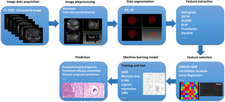
Recently, radiomic texture quantification of tumors has received much attention from radiologists, scientists, and stakeholders because several results have shown the feasibility of using the technique to diagnose and manage oncological conditions. In patients with hepatocellular carcinoma, radiomics has been applied in all stages of tumor evaluation, including diagnosis and characterization of the genotypic behavior of the tumor, monitoring of treatment responses and prediction of various clinical endpoints. It is also useful in selecting suitable candidates for specific treatment strategies. However, the clinical validation of hepatocellular carcinoma radiomics is limited by challenges in imaging protocol and data acquisition parameters, challenges in segmentation techniques, dimensionality reduction, and modeling methods. Identification of the best segmentation and optimal modeling methods, as well as texture features most stable to imaging protocol variability would go a long way in harmonizing HCC radiomics for personalized patient care. This article reviews the process of HCC radiomics, its clinical applications, associated challenges, and current optimization strategies.
近年来,肿瘤的放射组学纹理定量分析受到放射科医生、科学家和利益相关者的广泛关注,因为多项研究结果表明,该技术在诊断和管理肿瘤疾病方面具有可行性。在肝细胞癌患者中,放射组学已应用于肿瘤评估的各个阶段,包括诊断和肿瘤基因型行为的特征分析、治疗反应监测以及各种临床终点的预测。它在选择特定治疗策略的合适患者方面也很有用。然而,肝细胞癌放射组学的临床验证受到成像协议和数据采集参数、分割技术、降维和建模方法方面的挑战所限制。确定最佳的分割和最优的建模方法,以及对成像协议变化最稳定的纹理特征,将极大地促进 HCC 放射组学的个性化患者护理。本文综述了 HCC 放射组学的发展过程、临床应用、相关挑战以及目前的优化策略。