Department of Chemical and Biomolecular Engineering, North Carolina State University, Raleigh, NC, 27695, USA.
Department of Mathematics, North Carolina State University, Raleigh, NC, 27695, USA.
BMC Biol. 2020 Sep 23;18(1):130. doi: 10.1186/s12915-020-00861-w.
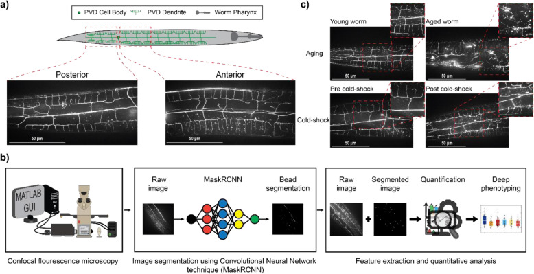
Access to quantitative information is crucial to obtain a deeper understanding of biological systems. In addition to being low-throughput, traditional image-based analysis is mostly limited to error-prone qualitative or semi-quantitative assessment of phenotypes, particularly for complex subcellular morphologies. The PVD neuron in Caenorhabditis elegans, which is responsible for harsh touch and thermosensation, undergoes structural degeneration as nematodes age characterized by the appearance of dendritic protrusions. Analysis of these neurodegenerative patterns is labor-intensive and limited to qualitative assessment.
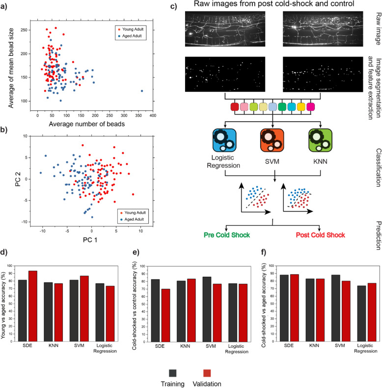
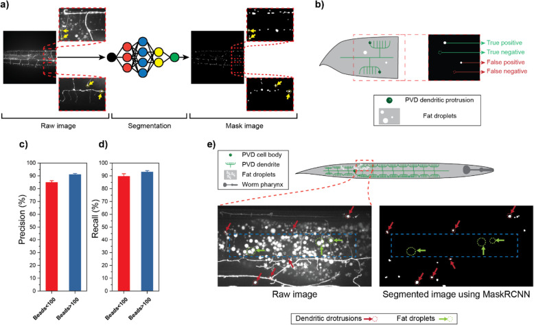
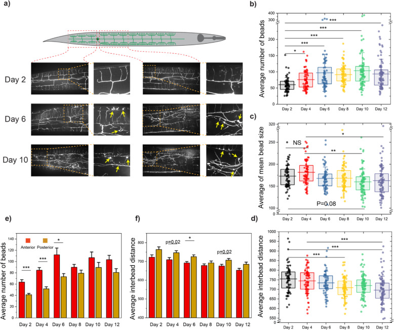
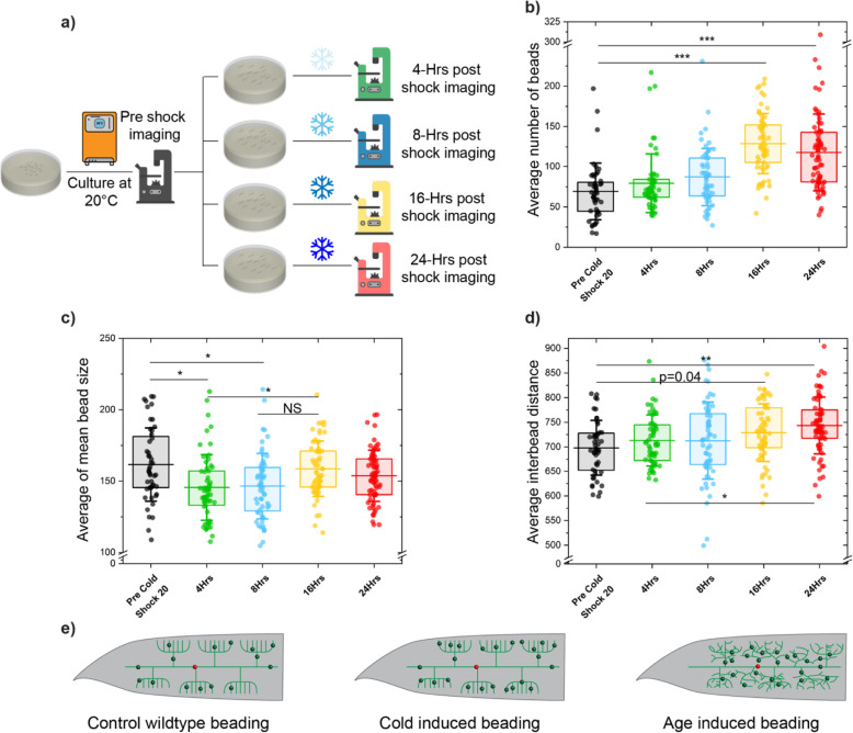
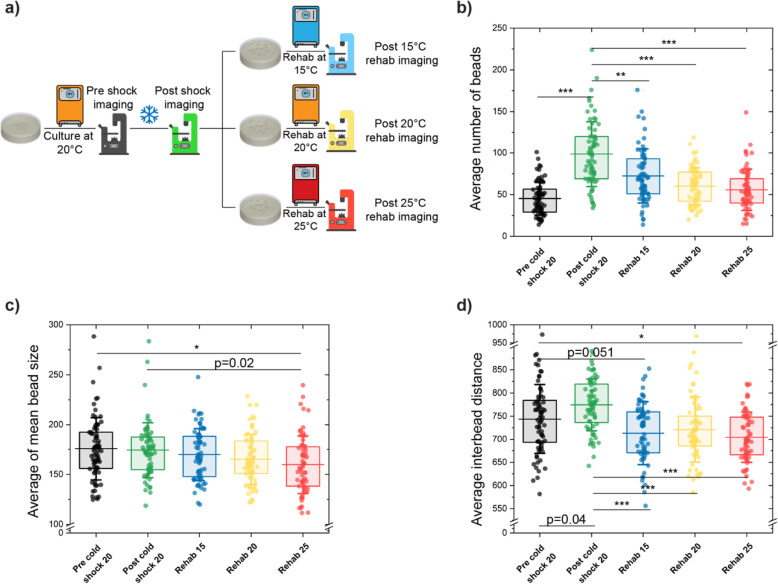
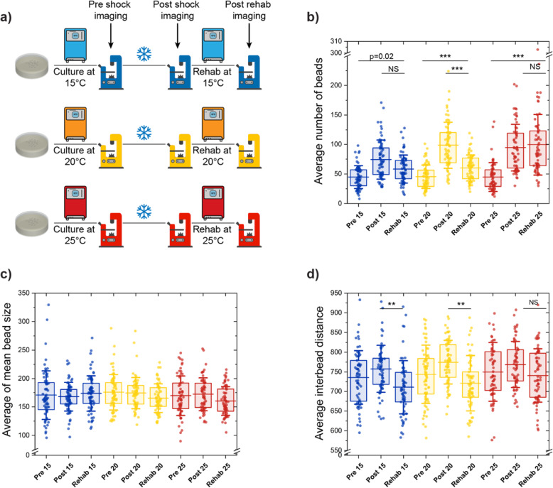
In this work, we apply deep learning to perform quantitative image-based analysis of complex neurodegeneration patterns exhibited by the PVD neuron in C. elegans. We apply a convolutional neural network algorithm (Mask R-CNN) to identify neurodegenerative subcellular protrusions that appear after cold-shock or as a result of aging. A multiparametric phenotypic profile captures the unique morphological changes induced by each perturbation. We identify that acute cold-shock-induced neurodegeneration is reversible and depends on rearing temperature and, importantly, that aging and cold-shock induce distinct neuronal beading patterns.
The results of this work indicate that implementing deep learning for challenging image segmentation of PVD neurodegeneration enables quantitatively tracking subtle morphological changes in an unbiased manner. This analysis revealed that distinct patterns of morphological alteration are induced by aging and cold-shock, suggesting different mechanisms at play. This approach can be used to identify the molecular components involved in orchestrating neurodegeneration and to characterize the effect of other stressors on PVD degeneration.
获取定量信息对于深入了解生物系统至关重要。除了通量低之外,传统的基于图像的分析方法大多仅限于对表型进行易错的定性或半定量评估,特别是对于复杂的亚细胞形态。秀丽隐杆线虫中的 PVD 神经元负责苛刻的触摸和热感觉,随着线虫年龄的增长,其结构会退化,表现为树突状突起的出现。这些神经退行性模式的分析既耗费劳力,又仅限于定性评估。
在这项工作中,我们应用深度学习对秀丽隐杆线虫 PVD 神经元表现出的复杂神经退行性模式进行定量的基于图像的分析。我们应用卷积神经网络算法(Mask R-CNN)来识别冷休克或衰老后出现的神经退行性亚细胞突起。多参数表型特征捕获了每种干扰诱导的独特形态变化。我们发现急性冷休克诱导的神经退行性变是可逆的,并且依赖于培养温度,重要的是,衰老和冷休克诱导了不同的神经元珠状模式。
这项工作的结果表明,对于 PVD 神经退行性变具有挑战性的图像分割实施深度学习可以实现以无偏方式定量跟踪微妙的形态变化。这种分析表明,衰老和冷休克诱导了不同的形态改变模式,表明存在不同的作用机制。这种方法可用于鉴定参与神经退行性变的分子成分,并描述其他应激源对 PVD 变性的影响。