Gao Xiangyu, Qiao Xiaona, Xing Xiaoxia, Huang Jinya, Qian Jiali, Wang Yi, Zhang Yawen, Zhang Xi, Li Miao, Cui Jiefeng, Yang Yehong
Department of Endocrinology, Huashan Hospital, Fudan University, Shanghai, China.
Liver Cancer Institute, Zhongshan Hospital, Fudan University & Key Laboratory of Carcinogenesis and Cancer Invasion, Ministry of Education, Shanghai, China.
Front Oncol. 2020 Aug 19;10:1563. doi: 10.3389/fonc.2020.01563. eCollection 2020.
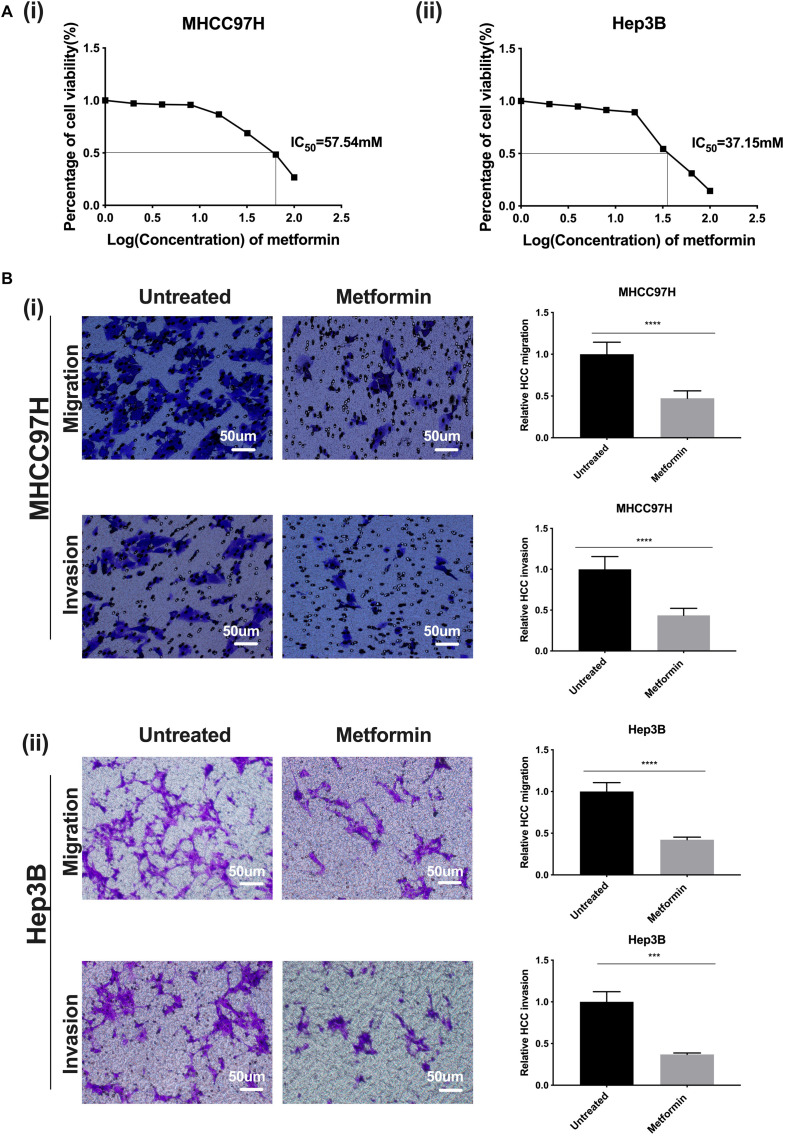
Metformin, a traditional first-line anti-hyperglycemic agent for diabetes, recently exhibits better antitumor effect in hepatocellular carcinoma (HCC). However, its resistance and tolerance mechanism in HCC remains largely unknown. Here, we investigated whether increased matrix stiffness attenuated the intervention effects of metformin on HCC invasion and metastasis, and explored its underlying molecular mechanism.
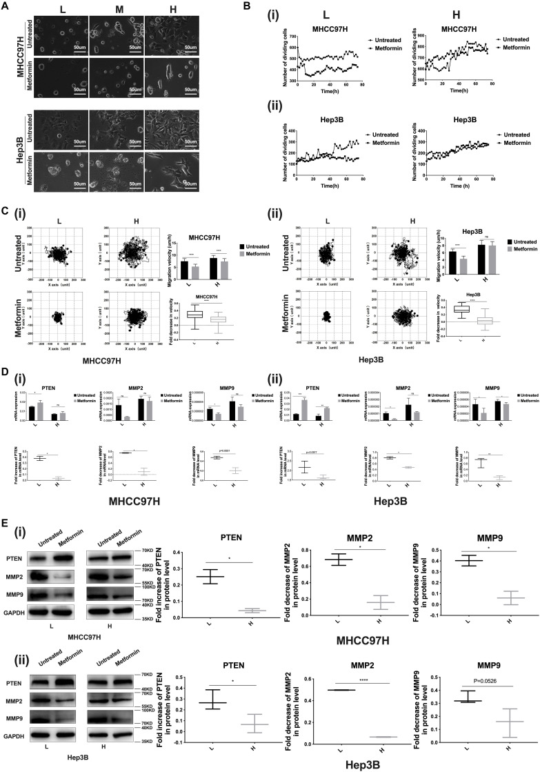
FN-coated gel substrates with 6, 10, and 16 kPa, which simulated the stiffness of normal, fibrotic, and cirrhotic liver tissues respectively, were established to evaluate matrix stiffness-mediated effects on HCC cells. Alterations in morphology, proliferation, motility, and invasive/metastatic-associated genes (PTEN, MMP2, MMP9) of HCC cells grown on different-stiffness substrates were comparatively analyzed before and after metformin intervention. Subsequently, the underlying molecular mechanism by which higher matrix stiffness attenuates antitumor effects of metformin in HCC was further elucidated.
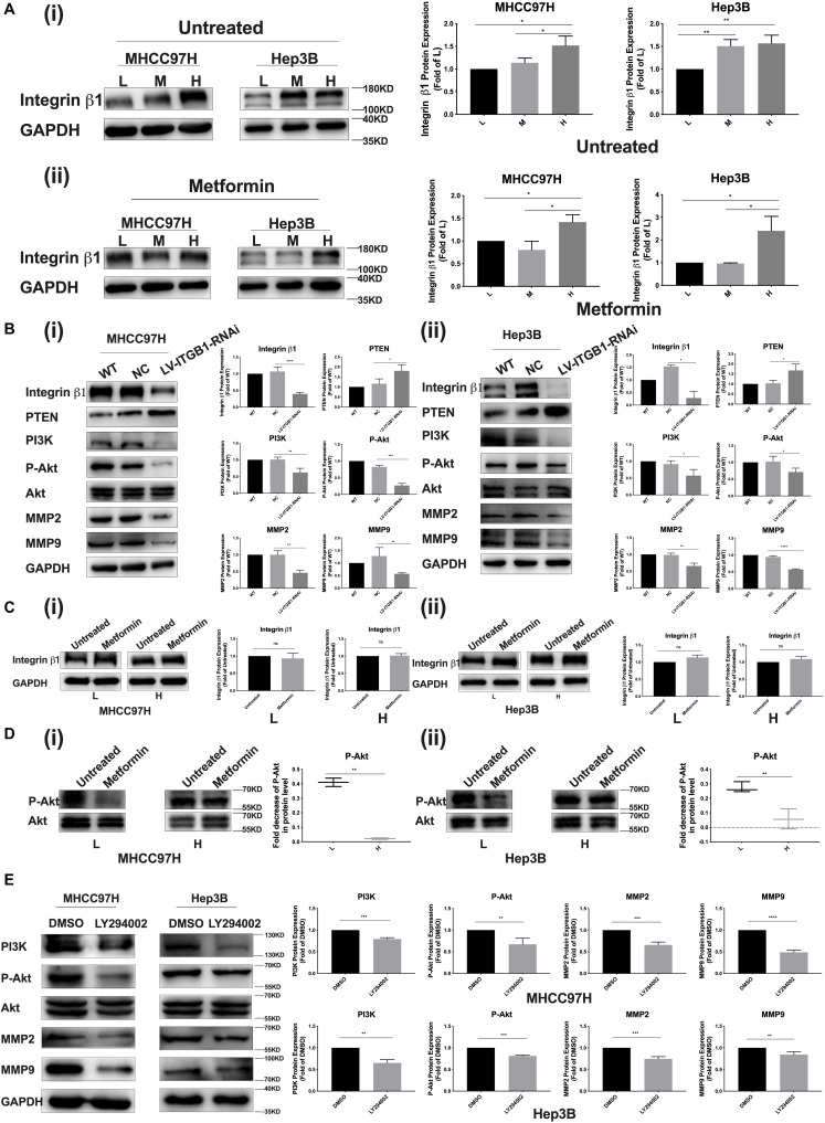
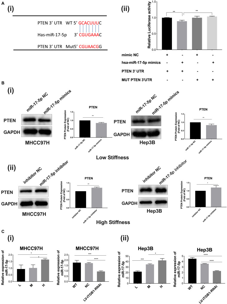
Metformin significantly inhibited proliferation, migration, and invasion of HCC cells. Compared with the controls on lower-stiffness substrate, HCC cells grown on higher-stiffness substrate exhibited an obvious resistance to intervention effects of metformin on proliferation, migration, invasion and metastasis. High stiffness stimulation significantly activated the miR-17-5p/PTEN/PI3K/Akt signaling pathway in HCC cells via integrin β1 and in turn resulted in MMP2 and MMP9 upregulation. Meanwhile, integrin β1 knockdown or PI3K inhibitor partially reversed the activation of the above signaling molecules. For HCC cells grown on the same-stiffness substrate, metformin remarkably upregulated PTEN expression and suppressed the activation of the PI3K/Akt/MMP pathway, but no effect on integrin β1 expression. Importantly, the increase in fold of PTEN expression and decrease in folds of Akt phosphorylation level and MMP2 and MMP9 expressions in the treated HCC cells with metformin on 16-kPa stiffness substrate were evidently weakened compared with those in the controls on the 6-kPa stiffness substrate.
Increased matrix stiffness significantly attenuates the inhibitory effect of metformin on HCC invasion and metastasis, and a common pathway of PTEN/PI3K/Akt/MMPs activated by mechanical stiffness signal and inactivated by metformin contributes to matrix stiffness-caused metformin resistance. To the best of our knowledge, this is the first report to clarify the mechanism of metformin intervention resistance from the perspective of tumor biophysical microenvironment.
二甲双胍作为传统的糖尿病一线降糖药物,近年来在肝细胞癌(HCC)中显示出较好的抗肿瘤作用。然而,其在HCC中的耐药及耐受机制仍不清楚。在此,我们研究了增加的基质硬度是否会减弱二甲双胍对HCC侵袭和转移的干预作用,并探讨其潜在的分子机制。
制备分别模拟正常、纤维化和肝硬化肝组织硬度的6、10和16 kPa纤维连接蛋白包被的凝胶基质,以评估基质硬度介导的对HCC细胞的影响。对在不同硬度基质上生长的HCC细胞在二甲双胍干预前后的形态、增殖、迁移以及侵袭/转移相关基因(PTEN、MMP2、MMP9)的变化进行比较分析。随后,进一步阐明更高的基质硬度减弱二甲双胍对HCC抗肿瘤作用的潜在分子机制。
二甲双胍显著抑制HCC细胞的增殖、迁移和侵袭。与生长在较低硬度基质上的对照细胞相比,生长在较高硬度基质上的HCC细胞对二甲双胍干预增殖、迁移、侵袭和转移的效果表现出明显抗性。高硬度刺激通过整合素β1显著激活HCC细胞中的miR-17-5p/PTEN/PI3K/Akt信号通路,进而导致MMP2和MMP9上调。同时,整合素β1敲低或PI3K抑制剂可部分逆转上述信号分子的激活。对于生长在相同硬度基质上的HCC细胞,二甲双胍显著上调PTEN表达并抑制PI3K/Akt/MMP通路的激活,但对整合素β1表达无影响。重要的是,与6 kPa硬度基质上的对照细胞相比,在16 kPa硬度基质上用二甲双胍处理的HCC细胞中PTEN表达倍数的增加以及Akt磷酸化水平和MMP2及MMP9表达倍数的降低明显减弱。
增加的基质硬度显著减弱二甲双胍对HCC侵袭和转移的抑制作用,由机械硬度信号激活并被二甲双胍失活的PTEN/PI3K/Akt/MMPs共同通路导致了基质硬度引起的二甲双胍耐药。据我们所知,这是第一份从肿瘤生物物理微环境角度阐明二甲双胍干预耐药机制的报告。