• 文献检索
  • 文档翻译
  • 深度研究
  • 学术资讯
  • Suppr Zotero 插件Zotero 插件
  • 邀请有礼
  • 套餐&价格
  • 历史记录
应用&插件
Suppr Zotero 插件Zotero 插件浏览器插件Mac 客户端Windows 客户端微信小程序
定价
高级版会员购买积分包购买API积分包
服务
文献检索文档翻译深度研究API 文档MCP 服务
关于我们
关于 Suppr公司介绍联系我们用户协议隐私条款
关注我们

Suppr 超能文献

核心技术专利:CN118964589B侵权必究
粤ICP备2023148730 号-1Suppr @ 2026

文献检索

告别复杂PubMed语法,用中文像聊天一样搜索,搜遍4000万医学文献。AI智能推荐,让科研检索更轻松。

立即免费搜索

文件翻译

保留排版,准确专业,支持PDF/Word/PPT等文件格式,支持 12+语言互译。

免费翻译文档

深度研究

AI帮你快速写综述,25分钟生成高质量综述,智能提取关键信息,辅助科研写作。

立即免费体验

基于扩散峰度成像和T2加权成像的放射组学分析用于鉴别胰腺神经内分泌肿瘤与实性假乳头状肿瘤

Radiomics Analysis Based on Diffusion Kurtosis Imaging and T2 Weighted Imaging for Differentiation of Pancreatic Neuroendocrine Tumors From Solid Pseudopapillary Tumors.

作者信息

Shi Yan-Jie, Zhu Hai-Tao, Liu Yu-Liang, Wei Yi-Yuan, Qin Xiu-Bo, Zhang Xiao-Yan, Li Xiao-Ting, Sun Ying-Shi

机构信息

Department of Radiology, Key Laboratory of Carcinogenesis and Translational Research, Peking University Cancer Hospital, Beijing, China.

出版信息

Front Oncol. 2020 Aug 21;10:1624. doi: 10.3389/fonc.2020.01624. eCollection 2020.

DOI:10.3389/fonc.2020.01624
PMID:32974201
原文链接:https://pmc.ncbi.nlm.nih.gov/articles/PMC7473210/
Abstract

OBJECTIVE

To develop and validate a radiomics model of diffusion kurtosis imaging (DKI) and T2 weighted imaging for discriminating pancreatic neuroendocrine tumors (PNETs) from solid pseudopapillary tumors (SPTs).

MATERIALS AND METHODS

Sixty-six patients with histopathological confirmed PNETs ( = 31) and SPTs ( = 35) were enrolled in this study. ROIs of tumors were manually drawn on each slice at T2WI and DWI ( = 1,500 s/mm) from 3T MRI. Intraclass correlation coefficients were used to evaluate the interobserver agreement. Mean diffusivity (MD) and mean kurtosis (MK) were derived from DKI. The least absolute shrinkage and selection operator regression were used for feature selection.

RESULTS

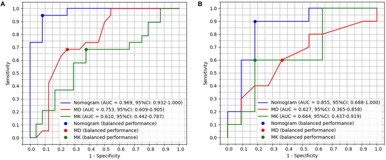
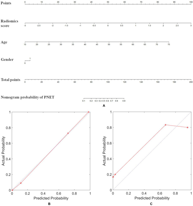
MD and MK had a moderate diagnostic performancewith the area under curve (AUC) of 0.71 and 0.65, respectively. A radiomics model, which incorporated sex and age of patients and radiomics signature of the tumor, showed excellent discrimination performance with AUC of 0.97 and 0.86 in the primary and validation cohort. Moreover, the new model had better diagnostic performance than that of MD ( = 0.023) and MK ( = 0.004), and showed excellent differentiation with a sensitivity of 95.00% and specificity of 91.67% in primary cohort, and the sensitivity of 90.91% and specificity of 81.82% in the validation cohort. The accuracy of radiomics analysis, radiologist 1, and radiologist 2 for diagnosing SPTs and PNETs were 92.42, 77.27, and 78.79%, respectively. The accuracy of radiomics analysis was significantly higher than that of subjective diagnosis ( < 0.05).

CONCLUSIONS

Radiomics model could improve the diagnostic accuracy of SPTs and PNETs and contribute to determining an appropriate treatment strategy for pancreatic tumors.

摘要

目的

建立并验证一种基于扩散峰度成像(DKI)和T2加权成像的影像组学模型,用于鉴别胰腺神经内分泌肿瘤(PNETs)和实性假乳头状肿瘤(SPTs)。

材料与方法

本研究纳入66例经组织病理学确诊的PNETs患者(n = 31)和SPTs患者(n = 35)。在3T MRI的T2WI和DWI(b = 1500 s/mm²)图像上,手动绘制肿瘤的感兴趣区(ROIs)。组内相关系数用于评估观察者间的一致性。从DKI中得出平均扩散率(MD)和平均峰度(MK)。采用最小绝对收缩和选择算子回归进行特征选择。

结果

MD和MK具有中等诊断性能,曲线下面积(AUC)分别为0.71和0.65。一个纳入患者性别、年龄和肿瘤影像组学特征的影像组学模型,在主要队列和验证队列中显示出优异的鉴别性能,AUC分别为0.97和0.86。此外,新模型的诊断性能优于MD(P = 0.023)和MK(P = 0.004),在主要队列中显示出优异的区分能力,敏感性为95.00%,特异性为91.67%,在验证队列中敏感性为90.91%,特异性为81.82%。影像组学分析、放射科医生1和放射科医生2诊断SPTs和PNETs的准确率分别为92.42%、77.27%和78.79%。影像组学分析的准确率显著高于主观诊断(P < 0.05)。

结论

影像组学模型可提高SPTs和PNETs的诊断准确性,并有助于确定胰腺肿瘤的合适治疗策略。

https://cdn.ncbi.nlm.nih.gov/pmc/blobs/e7a3/7473210/2b249c0a7c75/fonc-10-01624-g004.jpg
https://cdn.ncbi.nlm.nih.gov/pmc/blobs/e7a3/7473210/f8321d940a39/fonc-10-01624-g001.jpg
https://cdn.ncbi.nlm.nih.gov/pmc/blobs/e7a3/7473210/994bbe745c21/fonc-10-01624-g002.jpg
https://cdn.ncbi.nlm.nih.gov/pmc/blobs/e7a3/7473210/d17f442570a7/fonc-10-01624-g003.jpg
https://cdn.ncbi.nlm.nih.gov/pmc/blobs/e7a3/7473210/2b249c0a7c75/fonc-10-01624-g004.jpg

相似文献

1
Radiomics Analysis Based on Diffusion Kurtosis Imaging and T2 Weighted Imaging for Differentiation of Pancreatic Neuroendocrine Tumors From Solid Pseudopapillary Tumors.基于扩散峰度成像和T2加权成像的放射组学分析用于鉴别胰腺神经内分泌肿瘤与实性假乳头状肿瘤
Front Oncol. 2020 Aug 21;10:1624. doi: 10.3389/fonc.2020.01624. eCollection 2020.
2
Radiomics analysis from magnetic resonance imaging in predicting the grade of nonfunctioning pancreatic neuroendocrine tumors: a multicenter study.基于磁共振成像的影像组学分析预测无功能胰腺神经内分泌肿瘤分级:一项多中心研究。
Eur Radiol. 2024 Jan;34(1):90-102. doi: 10.1007/s00330-023-09957-7.
3
Non-gaussian models of 3-Tesla diffusion-weighted MRI for the differentiation of pancreatic ductal adenocarcinomas from neuroendocrine tumors and solid pseudopapillary neoplasms.3T 磁共振扩散加权成像的非高斯模型在胰腺导管腺癌与神经内分泌肿瘤和实性假乳头状肿瘤鉴别诊断中的应用。
Magn Reson Imaging. 2021 Nov;83:68-76. doi: 10.1016/j.mri.2021.07.006. Epub 2021 Jul 24.
4
MRI-based radiomics approach for differentiation of hypovascular non-functional pancreatic neuroendocrine tumors and solid pseudopapillary neoplasms of the pancreas.基于 MRI 的放射组学方法鉴别乏血供无功能胰腺神经内分泌肿瘤和胰腺实性假乳头状肿瘤。
BMC Med Imaging. 2021 Feb 23;21(1):36. doi: 10.1186/s12880-021-00563-x.
5
Radiomics Based on Multimodal MRI for the Differential Diagnosis of Benign and Malignant Breast Lesions.基于多模态磁共振成像的影像组学用于乳腺良恶性病变的鉴别诊断
J Magn Reson Imaging. 2020 Aug;52(2):596-607. doi: 10.1002/jmri.27098. Epub 2020 Feb 14.
6
T2-Weighted Image-Based Radiomics Signature for Discriminating Between Seminomas and Nonseminoma.基于T2加权图像的放射组学特征用于鉴别精原细胞瘤和非精原细胞瘤
Front Oncol. 2019 Nov 28;9:1330. doi: 10.3389/fonc.2019.01330. eCollection 2019.
7
Noncontrast Radiomics Approach for Predicting Grades of Nonfunctional Pancreatic Neuroendocrine Tumors.用于预测无功能胰腺神经内分泌肿瘤分级的非增强放射组学方法
J Magn Reson Imaging. 2020 Oct;52(4):1124-1136. doi: 10.1002/jmri.27176. Epub 2020 Apr 28.
8
Radiomics Analysis of Diffusion Kurtosis Imaging: Distinguishing Between Glioblastoma and Single Brain Metastasis.扩散峰度成像的放射组学分析:鉴别脑胶质母细胞瘤和单发脑转移瘤。
Acad Radiol. 2024 Mar;31(3):1036-1043. doi: 10.1016/j.acra.2023.07.023. Epub 2023 Sep 9.
9
Radiomics analysis of multiparametric MRI for the preoperative evaluation of pathological grade in bladder cancer tumors.多参数 MRI 影像组学分析在膀胱癌肿瘤术前病理分级评估中的应用。
Eur Radiol. 2019 Nov;29(11):6182-6190. doi: 10.1007/s00330-019-06222-8. Epub 2019 Apr 23.
10
A clinical-radiomics model incorporating T2-weighted and diffusion-weighted magnetic resonance images predicts the existence of lymphovascular invasion / perineural invasion in patients with colorectal cancer.纳入 T2 加权和弥散加权磁共振成像的临床放射组学模型预测结直肠癌患者的脉管侵犯/神经侵犯的存在。
Med Phys. 2021 Sep;48(9):4872-4882. doi: 10.1002/mp.15001. Epub 2021 Jul 21.

引用本文的文献

1
Advancements in Radiomics-Based AI for Pancreatic Ductal Adenocarcinoma.基于放射组学的人工智能在胰腺导管腺癌中的进展。
Bioengineering (Basel). 2025 Aug 6;12(8):849. doi: 10.3390/bioengineering12080849.
2
Prediction of solid pseudopapillary tumor invasiveness of the pancreas based on multiphase contrast-enhanced CT radiomics nomogram.基于多期增强CT影像组学列线图预测胰腺实性假乳头状瘤的侵袭性
Front Oncol. 2025 Apr 7;15:1513193. doi: 10.3389/fonc.2025.1513193. eCollection 2025.
3
Prognostic value of CT-based radiomics in grade 1-2 pancreatic neuroendocrine tumors.

本文引用的文献

1
Optimal b-values for diffusion kurtosis imaging of the liver and pancreas in MR examinations.肝脏和胰腺磁共振扩散峰度成像的最优 b 值。
Phys Med. 2019 Oct;66:119-123. doi: 10.1016/j.ejmp.2019.09.238. Epub 2019 Oct 7.
2
MRI Texture Analysis for Differentiating Nonfunctional Pancreatic Neuroendocrine Neoplasms From Solid Pseudopapillary Neoplasms of the Pancreas.MRI 纹理分析用于鉴别胰腺无功能性神经内分泌肿瘤与胰腺实性假乳头状肿瘤。
Acad Radiol. 2020 Jun;27(6):815-823. doi: 10.1016/j.acra.2019.07.012. Epub 2019 Aug 20.
3
Differentiation of atypical non-functional pancreatic neuroendocrine tumor and pancreatic ductal adenocarcinoma using CT based radiomics.
基于 CT 的影像组学对 1-2 级胰腺神经内分泌肿瘤的预后价值。
Cancer Imaging. 2024 Feb 23;24(1):28. doi: 10.1186/s40644-024-00673-z.
4
Prognostic Assessment of Gastropancreatic Neuroendocrine Neoplasm: Prospects and Limits of Radiomics.胃胰腺神经内分泌肿瘤的预后评估:影像组学的前景与局限
Diagnostics (Basel). 2023 Sep 7;13(18):2877. doi: 10.3390/diagnostics13182877.
5
Perioperative safety and prognosis following parenchyma-preserving surgery for solid pseudopapillary neoplasm of the pancreas.胰腺实性假乳头状瘤保留胰腺实质手术的围手术期安全性和预后。
World J Surg Oncol. 2023 Mar 31;21(1):119. doi: 10.1186/s12957-023-03003-y.
6
Can solid pseudopapillary neoplasm of the pancreas without degeneration be diagnosed with imaging? a comparison study of the solid component of solid pseudopapillary neoplasm, neuroendocrine neoplasm, and ductal adenocarcinoma.未发生退变的胰腺实性假乳头状肿瘤能否通过影像学诊断?实性假乳头状肿瘤、神经内分泌肿瘤及导管腺癌实性成分的比较研究
Abdom Radiol (NY). 2023 Mar;48(3):936-951. doi: 10.1007/s00261-023-03814-3. Epub 2023 Jan 28.
7
Pancreatic Mass Characterization Using IVIM-DKI MRI and Machine Learning-Based Multi-Parametric Texture Analysis.使用体素内不相干运动扩散峰度成像磁共振成像(IVIM-DKI MRI)和基于机器学习的多参数纹理分析对胰腺肿块进行特征描述
Bioengineering (Basel). 2023 Jan 8;10(1):83. doi: 10.3390/bioengineering10010083.
8
Age in combination with gender is a valuable parameter in differential diagnosis of solid pseudopapillary tumors and pancreatic neuroendocrine neoplasm.年龄与性别相结合是鉴别诊断实性假乳头状肿瘤和胰腺神经内分泌肿瘤的一个有价值的参数。
BMC Endocr Disord. 2022 Oct 21;22(1):255. doi: 10.1186/s12902-022-01164-7.
9
Advances in the imaging of gastroenteropancreatic neuroendocrine neoplasms.胃肠胰神经内分泌肿瘤的影像学进展。
World J Gastroenterol. 2022 Jul 14;28(26):3008-3026. doi: 10.3748/wjg.v28.i26.3008.
10
Comparison of the Differential Diagnostic Performance of Intravoxel Incoherent Motion Imaging and Diffusion Kurtosis Imaging in Malignant and Benign Thyroid Nodules.体素内不相干运动成像与扩散峰度成像在良恶性甲状腺结节鉴别诊断中的性能比较
Front Oncol. 2022 Jul 22;12:895972. doi: 10.3389/fonc.2022.895972. eCollection 2022.
基于 CT 影像组学鉴别不典型无功能胰腺神经内分泌肿瘤与胰腺导管腺癌
Eur J Radiol. 2019 Aug;117:102-111. doi: 10.1016/j.ejrad.2019.05.024. Epub 2019 May 31.
4
Diffusion kurtosis imaging and conventional diffusion weighted imaging to assess electrochemotherapy response in locally advanced pancreatic cancer.扩散峰度成像与常规扩散加权成像评估局部进展期胰腺癌电化学治疗反应的比较。
Radiol Oncol. 2019 Jan 19;53(1):15-24. doi: 10.2478/raon-2019-0004.
5
MR Imaging of Pancreatic Neuroendocrine Tumors.胰腺神经内分泌肿瘤的磁共振成像
Magn Reson Imaging Clin N Am. 2018 Aug;26(3):391-403. doi: 10.1016/j.mric.2018.03.010. Epub 2018 Jun 8.
6
Diffusion and perfusion MR parameters to assess preoperative short-course radiotherapy response in locally advanced rectal cancer: a comparative explorative study among Standardized Index of Shape by DCE-MRI, intravoxel incoherent motion- and diffusion kurtosis imaging-derived parameters.扩散和灌注磁共振参数评估局部进展期直肠癌术前短程放疗反应:DCE-MRI 标准化形状指数、体素内不相干运动和扩散峰度成像衍生参数的比较探索性研究。
Abdom Radiol (NY). 2019 Nov;44(11):3683-3700. doi: 10.1007/s00261-018-1801-z.
7
MR diffusion kurtosis imaging for cancer diagnosis: A meta-analysis of the diagnostic accuracy of quantitative kurtosis value and diffusion coefficient.磁共振扩散峰度成像在癌症诊断中的应用:定量峰度值与扩散系数诊断准确性的荟萃分析
Clin Imaging. 2018 Nov-Dec;52:44-56. doi: 10.1016/j.clinimag.2018.06.005. Epub 2018 Jun 9.
8
Building CT Radiomics Based Nomogram for Preoperative Esophageal Cancer Patients Lymph Node Metastasis Prediction.构建基于CT影像组学的列线图用于术前食管癌患者淋巴结转移预测
Transl Oncol. 2018 Jun;11(3):815-824. doi: 10.1016/j.tranon.2018.04.005. Epub 2018 May 1.
9
Diagnosis and treatment of solid-pseudopapillary tumors of the pancreas: A single institution experience with 97 cases.胰腺实性假乳头状瘤的诊断与治疗:单中心 97 例经验。
Pancreatology. 2018 Jun;18(4):415-419. doi: 10.1016/j.pan.2017.12.012. Epub 2017 Dec 24.
10
Radiomics Analysis for Evaluation of Pathological Complete Response to Neoadjuvant Chemoradiotherapy in Locally Advanced Rectal Cancer.放射组学分析评估局部晚期直肠癌新辅助放化疗的病理完全缓解。
Clin Cancer Res. 2017 Dec 1;23(23):7253-7262. doi: 10.1158/1078-0432.CCR-17-1038. Epub 2017 Sep 22.