Peeperkorn Sam, Meulemans Jeroen, Van Lierde Charlotte, Laenen Annouschka, Valstar Matthijs H, Balm A J M, Delaere Pierre, Vander Poorten Vincent
Otorhinolaryngology - Head and Neck Surgery, Leuven Cancer Institute, University Hospitals Leuven, Leuven, Belgium.
Section Head and Neck Oncology, Department of Oncology, KU Leuven, Leuven, Belgium.
Front Oncol. 2020 Aug 25;10:1535. doi: 10.3389/fonc.2020.01535. eCollection 2020.
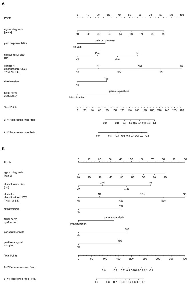
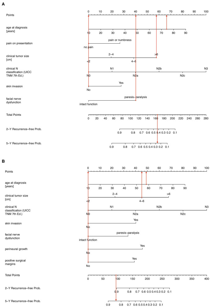
Salivary gland malignancies are rare tumors with a heterogenous histological and clinical appearance. Previously, we identified multiple prognostic factors in patients with parotid cancer and developed prognostic indices which have repeatedly been validated internationally, demonstrating their general applicability and lasting relevance. Recently, nomograms gained popularity as a prognostic tool. Thus, in this research we aimed to construct nomograms based on our previous validated prognostic models. Nomograms were constructed using the previously reported dataset of 168 patients with parotid cancer which was used to develop pre- and postoperative prognostic scores, PS1 and PS2, respectively. Concordance indices for PS1 and PS2 were previously estimated at 0.74 and 0.71, respectively, and are in line with other, widely accepted oncological nomograms. Pre- and postoperative nomograms predicting 2- and 5-year tumor recurrence-free survival probability are presented. All previously multivariately identified and validated prognostic factors, are incorporated (T size, N classification, pain, age at diagnosis, skin invasion, facial nerve dysfunction, perineural growth, and positive surgical margins). Examples of clinical application and interpretation are given. The presented prognostic nomograms for predicting 2- and 5-year tumor recurrence-free probability in patients with parotid cancer are powerful, user-friendly, visual tools and are based on internationally validated prognostic indices. They allow for a reliable prognostic assessment and result in a more individualized estimate of the risk for recurrence than the prognostic grouping based on PS1 and PS2. This facilitates assigning trial-patients to risk groups, and may assist in therapeutic decision making and determining appropriate follow-up intervals in clinical practice.
涎腺恶性肿瘤是一类罕见肿瘤,具有异质性的组织学和临床表现。此前,我们在腮腺癌患者中确定了多个预后因素,并制定了预后指数,这些指数已在国际上多次得到验证,证明了其普遍适用性和持久相关性。最近,列线图作为一种预后工具受到欢迎。因此,在本研究中,我们旨在基于我们之前经过验证的预后模型构建列线图。列线图是使用先前报道的168例腮腺癌患者的数据集构建的,该数据集分别用于制定术前和术后预后评分PS1和PS2。PS1和PS2的一致性指数此前分别估计为0.74和0.71,与其他广泛接受的肿瘤列线图一致。本文展示了预测2年和5年无肿瘤复发生存概率的术前和术后列线图。所有先前经多变量鉴定和验证的预后因素均被纳入(肿瘤大小、N分期、疼痛、诊断时年龄、皮肤侵犯、面神经功能障碍、神经周围生长和手术切缘阳性)。文中给出了临床应用和解读的示例。所展示的用于预测腮腺癌患者2年和5年无肿瘤复发概率的预后列线图是强大、用户友好的可视化工具,并且基于国际验证的预后指数。它们能够进行可靠的预后评估,与基于PS1和PS2的预后分组相比,能更个体化地估计复发风险。这有助于将试验患者分配到风险组,并可能有助于临床实践中的治疗决策和确定合适的随访间隔。